C. J. Gorter Center for High-Field MRI, Department of Radiology, Leiden University Medical Center, Leiden, The Netherlands.
Department of Radiology and Nuclear Medicine, Erasmus MC, University Medical Center Rotterdam, Rotterdam, The Netherlands.
NMR Biomed. 2022 May;35(5):e4653. doi: 10.1002/nbm.4653. Epub 2021 Nov 23.
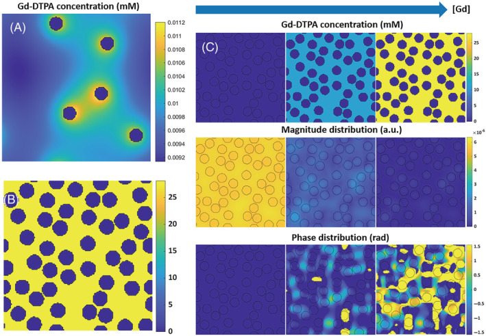
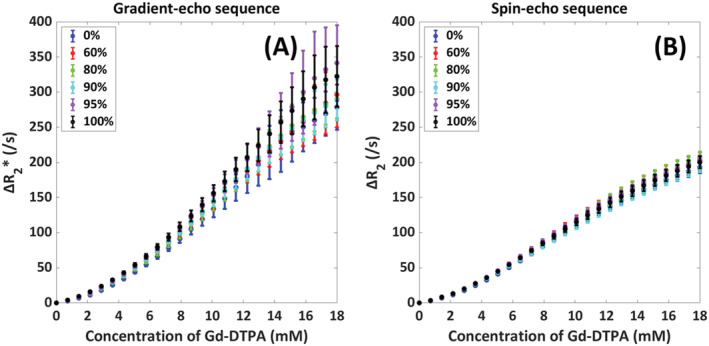
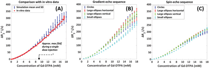
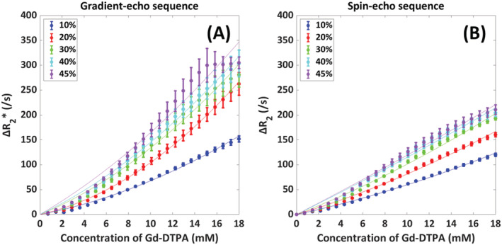
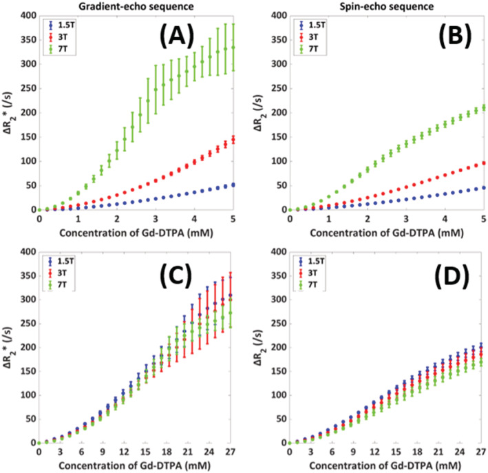
Dynamic susceptibility contrast (DSC) MRI is clinically used to measure brain perfusion by monitoring the dynamic passage of a bolus of contrast agent through the brain. For quantitative analysis of the DSC images, the arterial input function is required. It is known that the original assumption of a linear relation between the R relaxation and the arterial contrast agent concentration is invalid, although the exact relation is as of yet unknown. Studying this relation in vitro is time-consuming, because of the widespread variations in field strengths, MRI sequences, contrast agents, and physiological conditions. This study aims to simulate the R versus contrast concentration relation under varying physiological and technical conditions using an adapted version of an open-source simulation tool. The approach was validated with previously acquired data in human whole blood at 1.5 T by means of a gradient-echo sequence (proof-of-concept). Subsequently, the impact of hematocrit, field strength, and oxygen saturation on this relation was studied for both gradient-echo and spin-echo sequences. The results show that for both gradient-echo and spin-echo sequences, the relaxivity increases with hematocrit and field strength, while the hematocrit dependency was nonlinear for both types of MRI sequences. By contrast, oxygen saturation has only a minor effect. In conclusion, the simulation setup has proven to be an efficient method to rapidly calibrate and estimate the relation between R and gadolinium concentration in whole blood. This knowledge will be useful in future clinical work to more accurately retrieve quantitative information on brain perfusion.
动态对比磁共振成像(DSC MRI)被临床用于通过监测造影剂团块在大脑中的动态通过来测量脑灌注。为了对 DSC 图像进行定量分析,需要动脉输入函数。尽管确切的关系尚不清楚,但众所周知,R 弛豫与动脉造影剂浓度之间的线性关系的原始假设是无效的。由于场强、MRI 序列、造影剂和生理条件的广泛变化,在体外研究这种关系非常耗时。本研究旨在使用经过改编的开源模拟工具,模拟在不同生理和技术条件下 R 与对比浓度之间的关系。通过在 1.5 T 下使用梯度回波序列对人体全血进行的先前采集的数据进行了验证(概念验证)。随后,研究了梯度回波和自旋回波序列中血细胞比容、场强和氧饱和度对这种关系的影响。结果表明,对于梯度回波和自旋回波序列,弛豫率随血细胞比容和场强的增加而增加,而对于两种类型的 MRI 序列,血细胞比容的依赖性是非线性的。相比之下,氧饱和度的影响较小。总之,模拟设置已被证明是一种快速校准和估计全血中 R 与钆浓度之间关系的有效方法。这一知识将有助于未来的临床工作,更准确地获取脑灌注的定量信息。