Touet Amadeo, Schmiedt Yannick, Köller Jessica, Prangenberg Christian, Cucchi Davide, Welle Kristian, Endler Christoph, Scheidt Sebastian
Clinic for Orthopedics and Trauma Surgery, University Hospital of Bonn, 53127 Bonn, Germany.
Clinic for Diagnostic and Interventional Radiology, University Hospital of Bonn, 53127 Bonn, Germany.
J Clin Med. 2024 Dec 2;13(23):7332. doi: 10.3390/jcm13237332.
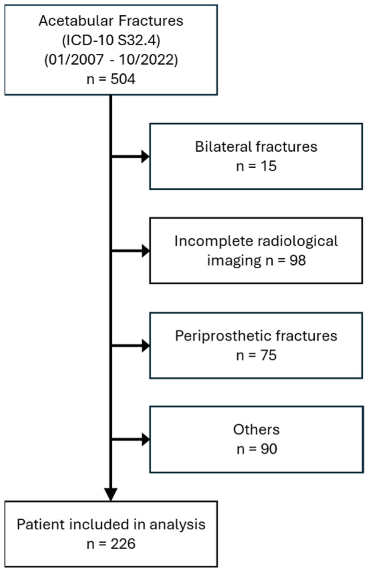
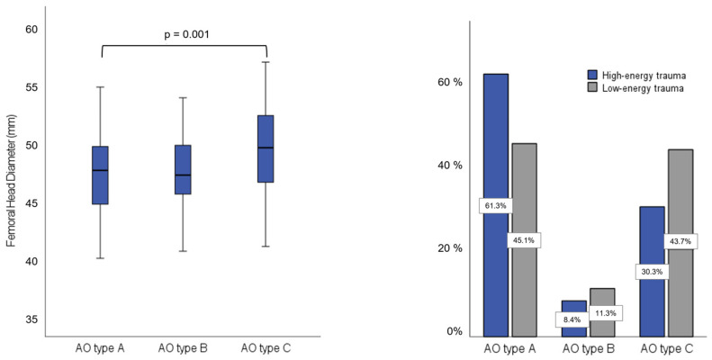
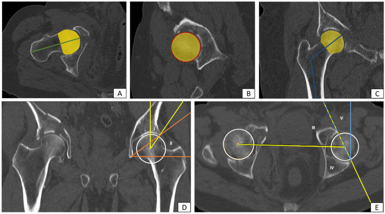
: Acetabular fractures continue to pose a major challenge in clinical practice, not least because of the growing geriatric population. While the influence of the force vectors on fracture formation is well established, the impact of anatomical factors on fracture morphology remains poorly understood. The aim of this study was to investigate patient-specific hip joint geometry, identify structural risk factors and correlate these with the resulting fracture patterns. : This retrospective cohort analysis included 226 patients (Mdn age = 58 yrs.) with acetabular fracture categorized by Judet/Letournel and the AO/OTA classification. Computed tomography (CT) datasets of the injured and contralateral sides were analyzed using multiplanar reconstruction. Parameters included modified center-edge (CE) angle (Wiberg), rotation angles (Ullmann and Anda), acetabular sector angle (Anda), true caput-collum-diaphyseal (CCD) angle, femoral head diameter and volume, as well as femoral neck length, circumference, and diameter. In addition, intrarater reliability within a subcohort was assessed for the metric measurements and inter-rater analysis for the classification of the entire sample. : The primary analysis showed direct effects of femoral head diameter, femoral neck length and femoral head size on the fracture type according to AO/OTA (type A/B/C), whereby this effect was particularly seen between type A and type C fractures ( = 0.001). Ordinal regression identified femoral head diameter as the only significant predictor ( = 0.02), with a 25% increased likelihood of complex fractures per unit of change. Low-energy trauma doubled the risk of severe fractures. Specific findings include a higher acetabular anteversion in anterior column fractures. Age correlated positively with the cause of injury and fracture type. The inter-rater reliability for fracture classification was excellent, as was the intrarater reliability of the measurements. : This study suggests that anatomical factors, particularly proximal femoral geometry, have an impact on acetabular fracture morphology-in addition to factors such as trauma type and patient demographics.
髋臼骨折在临床实践中仍然是一项重大挑战,尤其是考虑到老年人口的不断增加。虽然力向量对骨折形成的影响已得到充分证实,但解剖因素对骨折形态的影响仍知之甚少。本研究的目的是调查特定患者的髋关节几何形状,确定结构危险因素,并将这些因素与由此产生的骨折类型相关联。
这项回顾性队列分析纳入了226例髋臼骨折患者(年龄中位数 = 58岁),根据Judet/Letournel和AO/OTA分类进行分类。使用多平面重建分析受伤侧和对侧的计算机断层扫描(CT)数据集。参数包括改良中心边缘(CE)角(Wiberg)、旋转角(Ullmann和Anda)、髋臼扇形角(Anda)、真头颈骨干(CCD)角、股骨头直径和体积,以及股骨颈长度、周长和直径。此外,对一个亚组内的测量进行了评分者内信度评估,并对整个样本的分类进行了评分者间分析。
初步分析表明,根据AO/OTA分类,股骨头直径、股骨颈长度和股骨头大小对骨折类型有直接影响,其中这种影响在A型和C型骨折之间尤为明显(P = 0.001)。有序回归确定股骨头直径是唯一的显著预测因素(P = 0.02),每单位变化复杂骨折的可能性增加25%。低能量创伤使严重骨折的风险增加一倍。具体发现包括前柱骨折中髋臼前倾角较高。年龄与损伤原因和骨折类型呈正相关。骨折分类的评分者间信度极佳,测量的评分者内信度也是如此。
本研究表明,除创伤类型和患者人口统计学等因素外,解剖因素,特别是股骨近端几何形状,对髋臼骨折形态有影响。