Soto Ricardo F, Godoy Sebastián E
Department of Electrical Engineering, Universidad de Concepción, Edmundo Larenas 219, Concepción, 4030000, Biobío, Chile.
Heliyon. 2024 Nov 26;10(23):e40608. doi: 10.1016/j.heliyon.2024.e40608. eCollection 2024 Dec 15.
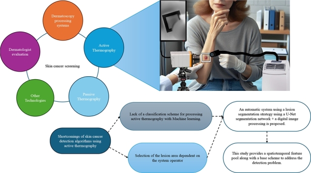
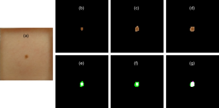
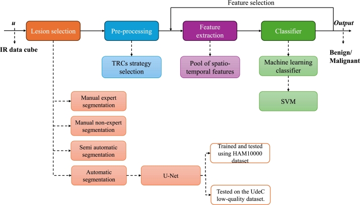
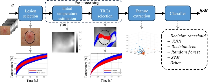
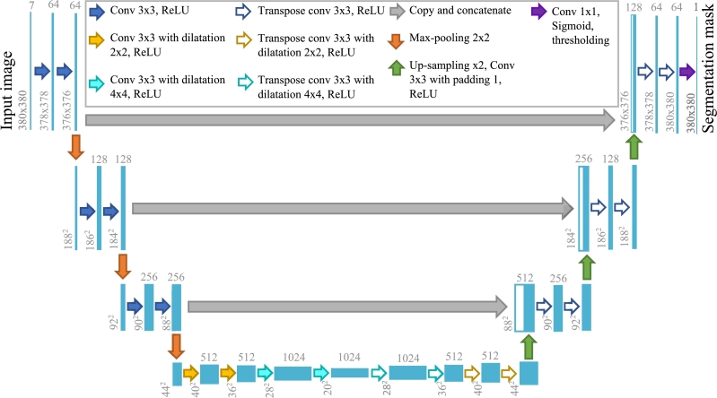
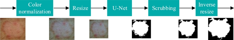
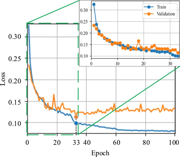
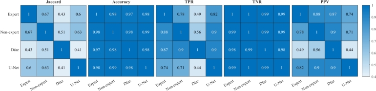
Skin cancer is a growing global concern, with cases steadily rising. Typically, malignant moles are identified through visual inspection, using dermatoscopy and patient history. Active thermography has emerged as an effective method to distinguish between malignant and benign lesions. Our previous research showed that spatio-temporal features can be extracted from suspicious lesions to accurately determine malignancy, which was applied in a distance-based classifier. In this study, we build on that foundation by introducing a set of novel spatial and temporal features that enhance classification accuracy and can be integrated into any machine learning approach. These features were implemented in a support-vector machine classifier to detect malignancy. Notably, our method addresses a common limitation in existing approaches-manual lesion selection-by automating the process using a U-Net convolutional neural network. We validated our system by comparing U-Net's performance with expert dermatologist segmentations, achieving a 17% improvement in the Jaccard index over a semi-automatic algorithm. The detection algorithm relies on accurate lesion segmentation, and its performance was evaluated across four segmentation techniques. At an 85% sensitivity threshold, expert segmentation provided the highest specificity at 87.62%, while non-expert and U-Net segmentations achieved comparable results of 69.63% and 68.80%, respectively. Semi-automatic segmentation lagged behind at 64.45%. This automated detection system performs comparably to high-accuracy methods while offering a more standardized and efficient solution. The proposed automatic system achieves 3% higher accuracy compared to the ResNet152V2 network when processing low-quality images obtained in a clinical setting.
皮肤癌是一个日益引起全球关注的问题,病例数在稳步上升。通常,恶性痣是通过目视检查、使用皮肤镜检查和患者病史来识别的。主动热成像已成为区分恶性和良性病变的有效方法。我们之前的研究表明,可以从可疑病变中提取时空特征以准确确定恶性程度,并将其应用于基于距离的分类器中。在本研究中,我们在此基础上引入了一组新颖的空间和时间特征,这些特征提高了分类准确率,并且可以集成到任何机器学习方法中。这些特征在支持向量机分类器中实现以检测恶性程度。值得注意的是,我们的方法通过使用U-Net卷积神经网络自动化流程,解决了现有方法中的一个常见限制——手动病变选择。我们通过将U-Net的性能与皮肤科专家的分割结果进行比较来验证我们的系统,在Jaccard指数上比半自动算法提高了17%。检测算法依赖于准确的病变分割,并且其性能在四种分割技术上进行了评估。在85%的灵敏度阈值下,专家分割的特异性最高,为87.62%,而非专家和U-Net分割的特异性分别为69.63%和68.80%,结果相当。半自动分割的特异性为64.45%,落后于其他方法。这种自动检测系统的性能与高精度方法相当,同时提供了更标准化和高效的解决方案。当处理在临床环境中获得的低质量图像时,所提出的自动系统比ResNet152V2网络的准确率高3%。