eVida Research Laboratory, University of Deusto, Avda. Universidades 24, 48007, Bilbao, Spain.
Department of Electrical and Electronics Engineering, Universidad del Norte, Km.5 Vía Puerto Colombia, Barranquilla, Colombia.
BMC Med Imaging. 2021 Jan 6;21(1):6. doi: 10.1186/s12880-020-00534-8.
Melanoma has become more widespread over the past 30 years and early detection is a major factor in reducing mortality rates associated with this type of skin cancer. Therefore, having access to an automatic, reliable system that is able to detect the presence of melanoma via a dermatoscopic image of lesions and/or skin pigmentation can be a very useful tool in the area of medical diagnosis.
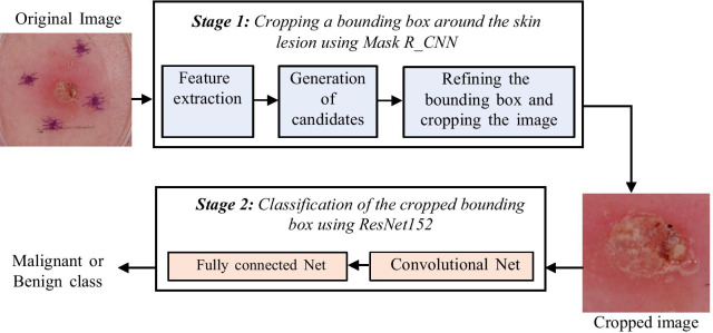
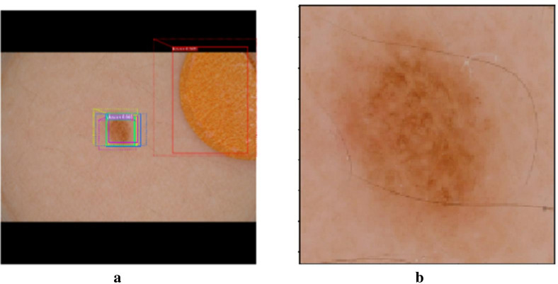
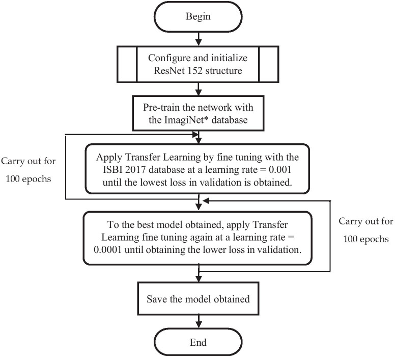
Among state-of-the-art methods used for automated or computer assisted medical diagnosis, attention should be drawn to Deep Learning based on Convolutional Neural Networks, wherewith segmentation, classification and detection systems for several diseases have been implemented. The method proposed in this paper involves an initial stage that automatically crops the region of interest within a dermatoscopic image using the Mask and Region-based Convolutional Neural Network technique, and a second stage based on a ResNet152 structure, which classifies lesions as either "benign" or "malignant".
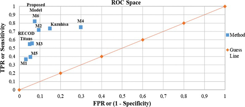
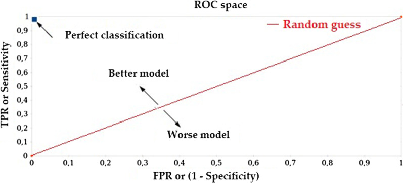
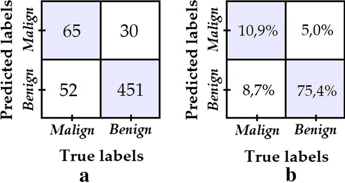
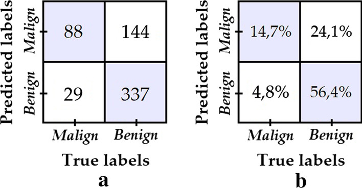
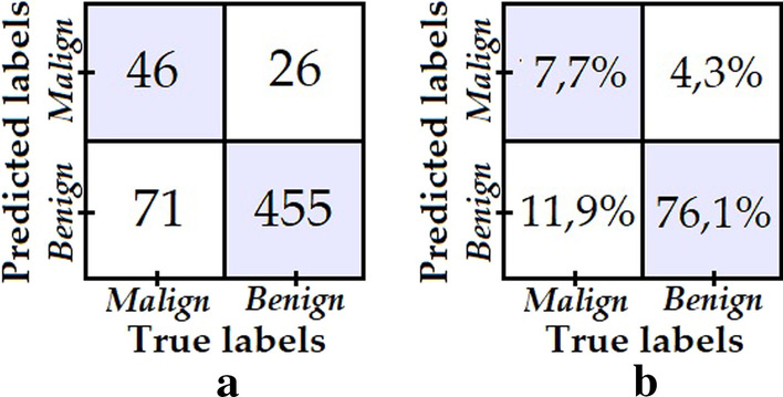
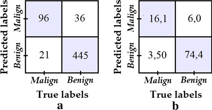
Training, validation and testing of the proposed model was carried out using the database associated to the challenge set out at the 2017 International Symposium on Biomedical Imaging. On the test data set, the proposed model achieves an increase in accuracy and balanced accuracy of 3.66% and 9.96%, respectively, with respect to the best accuracy and the best sensitivity/specificity ratio reported to date for melanoma detection in this challenge. Additionally, unlike previous models, the specificity and sensitivity achieve a high score (greater than 0.8) simultaneously, which indicates that the model is good for accurate discrimination between benign and malignant lesion, not biased towards any of those classes.
The results achieved with the proposed model suggest a significant improvement over the results obtained in the state of the art as far as performance of skin lesion classifiers (malignant/benign) is concerned.
在过去的 30 年中,黑色素瘤的发病率有所上升,早期发现是降低与这种皮肤癌相关死亡率的主要因素。因此,拥有一种能够通过病变和/或皮肤色素的皮肤镜图像自动、可靠地检测黑色素瘤存在的自动系统,可能是医学诊断领域非常有用的工具。
在用于自动或计算机辅助医学诊断的最新方法中,应注意基于卷积神经网络的深度学习,已经使用这种方法实现了几种疾病的分割、分类和检测系统。本文提出的方法涉及一个初始阶段,该阶段使用基于掩模和区域的卷积神经网络技术自动裁剪皮肤镜图像中的感兴趣区域,以及基于 ResNet152 结构的第二阶段,该阶段将病变分类为“良性”或“恶性”。
使用与 2017 年国际生物医学成像研讨会挑战赛相关的数据库对所提出的模型进行了训练、验证和测试。在测试数据集上,与迄今为止在该挑战赛中报告的最佳准确性和最佳灵敏度/特异性比相比,所提出的模型在准确性和平衡准确性方面分别提高了 3.66%和 9.96%。此外,与以前的模型不同,特异性和灵敏度同时达到了较高的分数(大于 0.8),这表明该模型非常适合准确区分良性和恶性病变,而不是偏向于任何一类病变。
所提出模型的结果表明,与皮肤病变分类器(良性/恶性)的最新技术相比,性能有了显著提高。