Jakimiuk Adam, Maintz Michaela, Müller-Gerbl Magdalena, Thieringer Florian Markus, Keller Marco, Guebeli Alissa, Honigmann Philipp
Department of Biomedical Engineering, Medical Additive Manufacturing Research Group (Swiss MAM), University of Basel, Allschwil, Switzerland.
Oral and Cranio-Maxillofacial Surgery, University Hospital Basel, Basel, Switzerland.
3D Print Med. 2024 Dec 18;10(1):42. doi: 10.1186/s41205-024-00240-z.
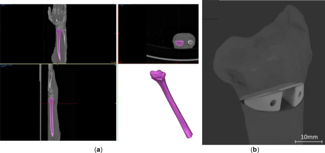
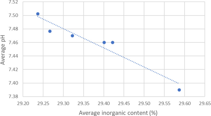
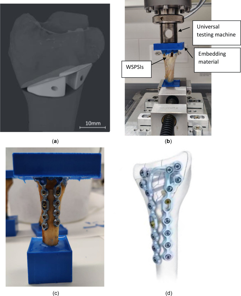
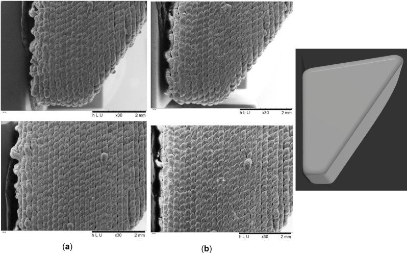
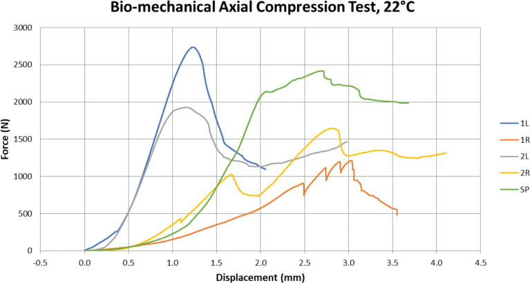
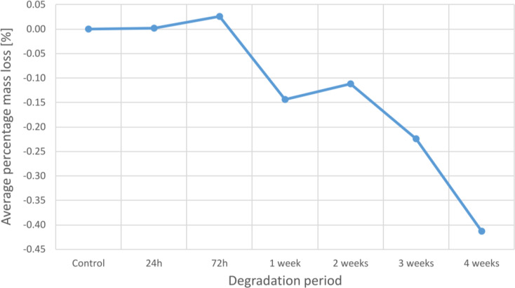
The most common surgical procedure to manage the malunion of the bones is corrective osteotomy. The current gold standard for securing the bone segments after osteotomy is the use of titanium plates and allografts which have disadvantages such as possible allergic reaction, additional operations such as extraction of the graft from other sites and removal operation. The utilization of resorbable materials presents an opportunity to mitigate these drawbacks but has not yet been thoroughly researched in the literature. This study assesses the viability of using biodegradable, 3D-printed patient-specific implants made of Poly(-L-lactide-co-D, L-lactide) (PLDLLA) and β-Tricalcium Phosphate (β-TCP) as an alternative material in an in-vitro biomechanical study involving ex vivo biomechanical compression testing, biodegradation testing, and calorimetric measurements. These implants possess a unique shape, resembling a wedge and are fixated as a connection between the osteotomised bone using resorbable screws. Following point-of-care virtual planning, bio-mechanical compressive tests with (n = 5) ex vivo radii equipped with PLDLLA/ β-TCP implants were performed to prove sufficient stability of the connection. All PLDLLA/ β-TCP implants withstood a compressive force of at least 1'211 N which exceeds the maximum force reported in literature in case of a fall from the height of one meter. Furthermore, the results showed a consistent surface chemistry and slow degradation rate. The outcomes are encouraging, establishing the groundwork for an innovative distal radius corrective osteotomy surgical method. However, further research is necessary to thoroughly evaluate the long-term biodegradability and mechanical efficacy of the implants.
治疗骨骼畸形愈合最常见的外科手术是截骨矫正术。截骨术后固定骨段的当前金标准是使用钛板和同种异体移植物,这些方法存在一些缺点,如可能的过敏反应、从其他部位取出移植物等额外手术以及移除手术。可吸收材料的应用为减轻这些缺点提供了机会,但在文献中尚未得到充分研究。本研究在一项体外生物力学研究中评估了使用由聚(-L-丙交酯-co-D,L-丙交酯)(PLDLLA)和β-磷酸三钙(β-TCP)制成的可生物降解、3D打印的患者特异性植入物作为替代材料的可行性,该研究包括离体生物力学压缩测试、生物降解测试和量热测量。这些植入物具有独特的形状,类似楔形,并使用可吸收螺钉作为截骨后的骨连接固定。在床旁虚拟规划之后,对(n = 5)个配备PLDLLA/β-TCP植入物的离体桡骨进行了生物力学压缩测试,以证明连接具有足够的稳定性。所有PLDLLA/β-TCP植入物都承受了至少1211 N的压缩力,超过了文献中报道的从一米高处跌落时的最大力。此外,结果显示表面化学性质一致且降解速率缓慢。这些结果令人鼓舞,为一种创新的桡骨远端截骨矫正手术方法奠定了基础。然而,需要进一步研究以全面评估植入物的长期生物降解性和机械效能。