Pitrone Pietro, Cacciola Agatino, Cattafi Antonino, Romeo Alessia Maria, Cracò Annalisa, Aricò Francesco Marcello, Migliaccio Nicola, Magnani Francesca, Bellone Italo Giuseppe, Caloggero Simona, Mastroeni Giampiero
Radiology Unit, "Papardo" Hospital, Messina, ME 98158, Italy.
Diagnostic and Interventional Radiology Unit, BIOMORF Department, University Hospital "Policlinico G. Martino", Messina, ME 98124, Italy.
Radiol Case Rep. 2024 Dec 3;20(2):1208-1210. doi: 10.1016/j.radcr.2024.11.025. eCollection 2025 Feb.
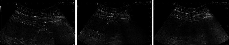
Multinodular steatosis represents a relatively uncommon manifestation of fatty liver disease (FLD). Co-morbidities such as metabolic syndrome or cirrhosis are often associated. Despite typical features of imaging (ultrasound, CT, and MRI), core biopsy sometimes remains the gold standard for diagnosis. We describe the case of a 57-year-old male patient with a long history of hepatic cirrhosis and a recent diagnosis of carcinoma of the tongue, successfully treated. Due to the occurrence of nausea, diarrhea and jaundice the patient is admitted to Our Hospital where ultrasound examination and contrast-enhanced CT are performed, showing global hypoechogenicity of the liver parenchyma with multiple hypo-attenuating lesions. To rule out metastatic lesions, contrast-enhanced CT of the thorax and cranium and gastroscopy and colonoscopy are performed, with no evidence of primary malignancy. Core biopsy reveals macro-vacuolar steatosis within a cirrhotic liver with regenerative aspects.
多结节性脂肪变性是脂肪性肝病(FLD)相对不常见的一种表现形式。常伴有代谢综合征或肝硬化等合并症。尽管有典型的影像学特征(超声、CT和MRI),但有时肝穿刺活检仍是诊断的金标准。我们描述了一名57岁男性患者的病例,该患者有长期肝硬化病史,近期诊断为舌癌,已成功接受治疗。由于出现恶心、腹泻和黄疸,该患者入住我院,进行了超声检查和增强CT,结果显示肝实质整体回声减低,伴有多个低密度病变。为排除转移性病变,进行了胸部和头颅增强CT以及胃镜和结肠镜检查,未发现原发性恶性肿瘤证据。肝穿刺活检显示肝硬化肝脏内有大泡性脂肪变性,并伴有再生表现。