Gorka Stephanie M, Jimmy Jagan, Koning Katherine, Phan K Luan, Rotstein Natalie, Hoang-Dang Bianca, Halavi Sabrina, Spivak Norman, Monti Martin M, Reggente Nicco, Bookheimer Susan Y, Kuhn Taylor P
Department of Psychiatry and Behavioral Health, Wexner Medical Center, The Ohio State University, Columbus, OH, United States.
Department of Psychiatry and Biobehavioral Sciences, The University of California, Los Angeles, Los Angeles, CA, United States.
Front Hum Neurosci. 2024 Dec 4;18:1486770. doi: 10.3389/fnhum.2024.1486770. eCollection 2024.
Low-intensity transcranial focused ultrasound (tFUS) is a brain stimulation approach that holds promise for the treatment of brain-based disorders. Studies in humans have shown that tFUS can successfully modulate perfusion in focal sonication targets, including the amygdala; however, limited research has explored how tFUS impacts large-scale neural networks.
The aim of the current study was to address this gap and examine changes in resting-state connectivity between large-scale network nodes using a randomized, double-blind, within-subjects crossover study design.
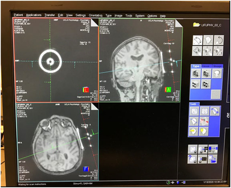
Healthy adults ( 18) completed two tFUS sessions, 14 days apart. Each session included tFUS of either the right amygdala or the left entorhinal cortex (ErC). The inclusion of two active targets allowed for within-subjects comparisons as a function of the locus of sonication. Resting-state functional magnetic resonance imaging was collected before and after each tFUS session.
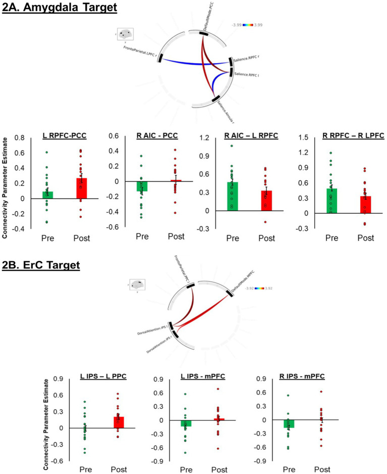
tFUS altered resting-state functional connectivity (rsFC) within and between rs-network nodes. Pre-to-post sonication of the right amygdala modulated connectivity within nodes of the salience network (SAN) and between nodes of the SAN and the default mode network (DMN) and frontoparietal network (FRP). A decrease in SAN to FPN connectivity was specific to the amygdala target. Pre-to-post sonication of the left ErC modulated connectivity between the dorsal attention network (DAN) and FPN and DMN. An increase in DAN to DMN connectivity was specific to the ErC target.
These preliminary findings may suggest that tFUS induces neuroplastic changes beyond the immediate sonication target. Additional studies are needed to determine the long-term stability of these effects.
低强度经颅聚焦超声(tFUS)是一种脑刺激方法,有望用于治疗脑部疾病。人体研究表明,tFUS能够成功调节局部超声照射靶点(包括杏仁核)的灌注;然而,关于tFUS如何影响大规模神经网络的研究却很有限。
本研究旨在填补这一空白,采用随机、双盲、受试者内交叉研究设计,研究大规模网络节点之间静息态连接性的变化。
健康成年人(≥18岁)完成两次tFUS治疗,间隔14天。每次治疗包括对右侧杏仁核或左侧内嗅皮层(ErC)进行tFUS照射。纳入两个活跃靶点,以便进行受试者内比较,作为超声照射部位的函数。在每次tFUS治疗前后采集静息态功能磁共振成像。
tFUS改变了静息态功能连接(rsFC),包括rs网络节点内部以及节点之间。右侧杏仁核超声照射前后调节了突显网络(SAN)节点内部以及SAN与默认模式网络(DMN)和额顶叶网络(FRP)节点之间的连接性。SAN与FPN连接性的降低是杏仁核靶点特有的。左侧ErC超声照射前后调节了背侧注意网络(DAN)与FPN和DMN之间的连接性。DAN与DMN连接性的增加是ErC靶点特有的。
这些初步研究结果可能表明,tFUS会在直接超声照射靶点之外诱导神经可塑性变化。需要进一步研究来确定这些效应的长期稳定性。