Wu Jiajun, Zhang Linyuan, Shen Chao, Wang Xiuhui, Zhou Xiaoxiao
Department of Orthopedics, Shanghai University of Medicine and Health Sciences Affiliated Zhoupu Hospital, 1500 Zhouyuan Road, Pudong New Area, Shanghai, 201318, China.
Department of Orthopedics, Jiangwan Hospital of Shanghai Hongkou District, 1878 Sichuan North Road Hongkou District, Shanghai, 200434, People's Republic of China.
BMC Musculoskelet Disord. 2024 Dec 19;25(1):1044. doi: 10.1186/s12891-024-08020-w.
Calcaneal fracture fixation remains a challenging procedure in orthopedics, with computational tools increasingly aiding in the optimization of preoperative planning. To compare the biomechanical stability of intramedullary fixation and locking plate fixation for Sanders II and III calcaneal fractures by three-dimensional (3D) finite element analysis and to provide a theoretical basis for clinical application.
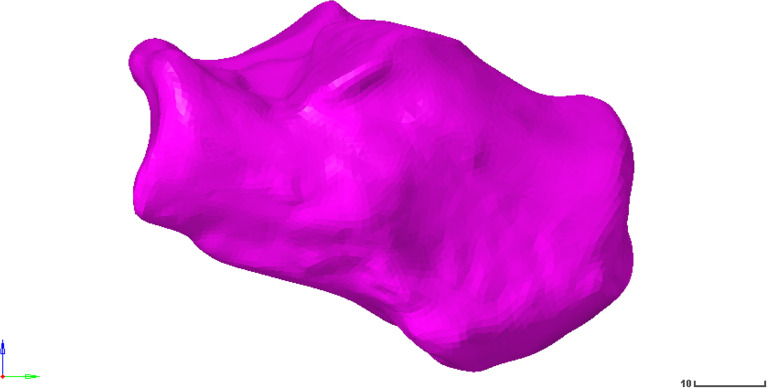
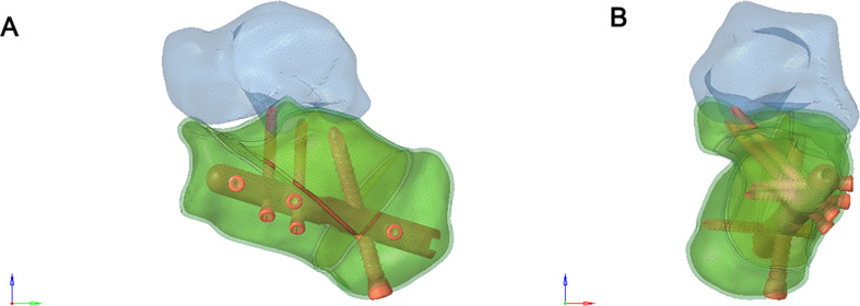
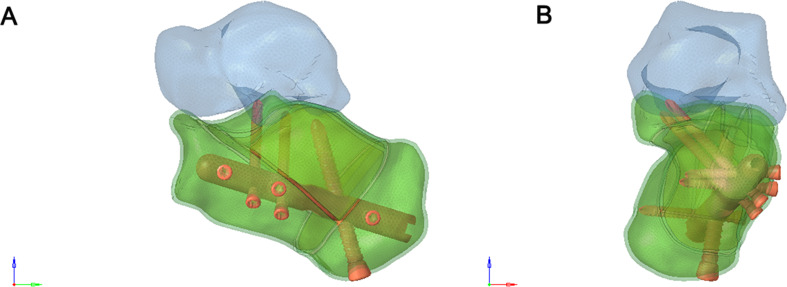
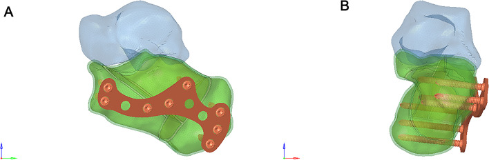
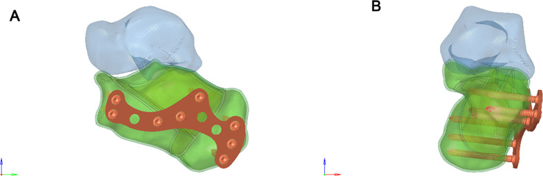
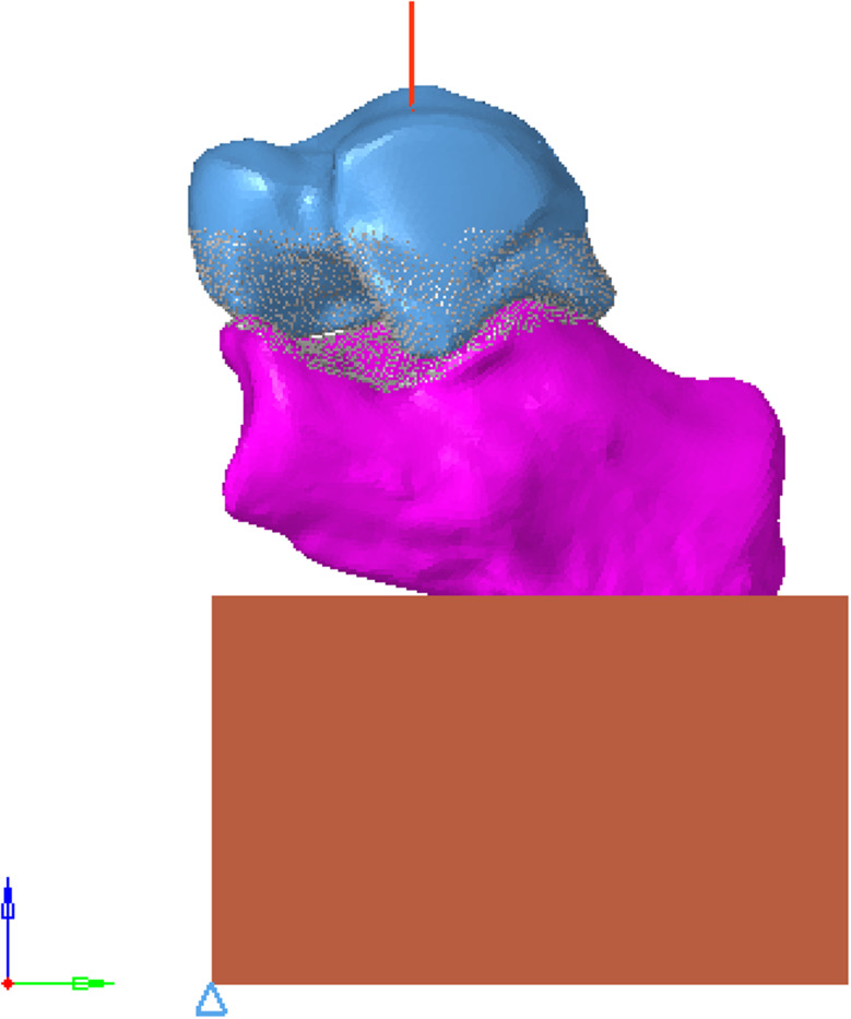
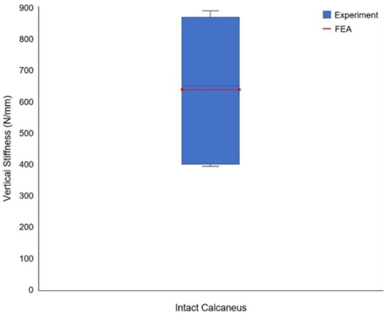
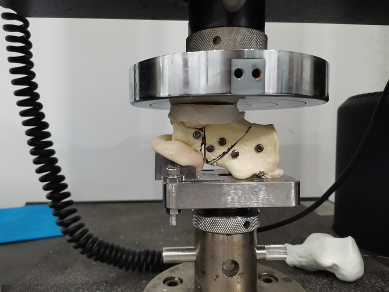
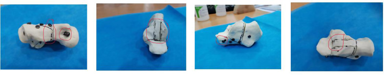
The Computed Tomography (CT) images were segmented using Mimics software (Materialise NV, Belgium) to identify the region of interest based on threshold segmentation. The 3D morphology was reconstructed using Mimics 10.01 software. Subsequently, Geomagic2012 software (3D Systems, USA) was employed to remove noise points, sharp corners, and scattered points, achieving a smooth surface map. This map was saved in Initial Graphics Exchange Specification (IGES) format and imported into Solidworks (Dassault Systèmes, France) for model assembly and volume model construction. A three-dimensional finite element model of Sanders II/III, calcaneal fractures with intramedullary fixation and locking plate fixation, was established and analyzed by linear finite element analysis. The forced displacement, stiffness, and stress distribution of the two fixation methods were calculated. In addition, the three-dimensional model was tested using a compression mechanical experiment.
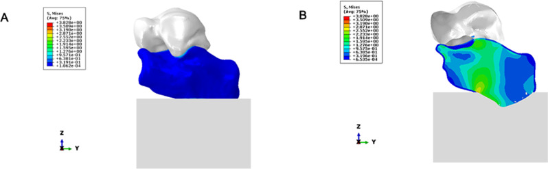
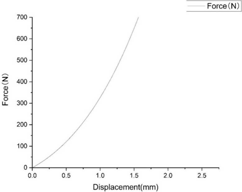
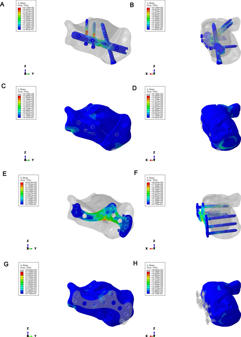
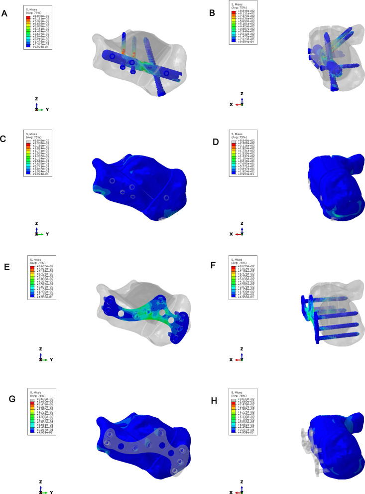
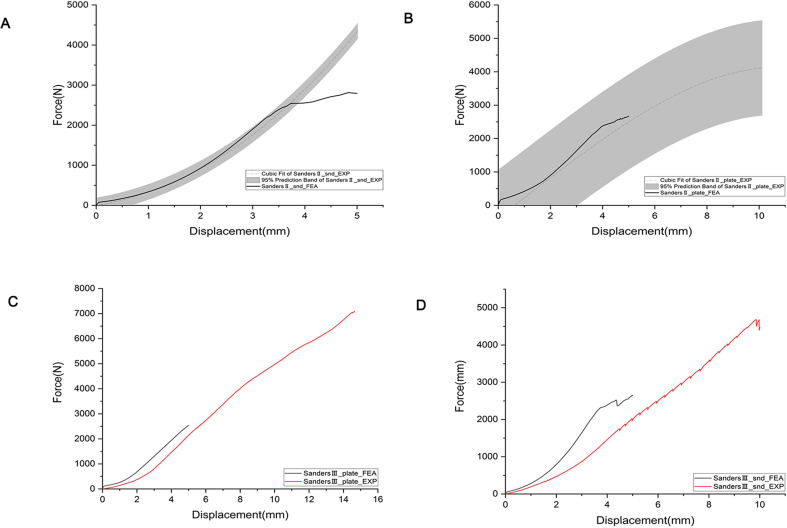
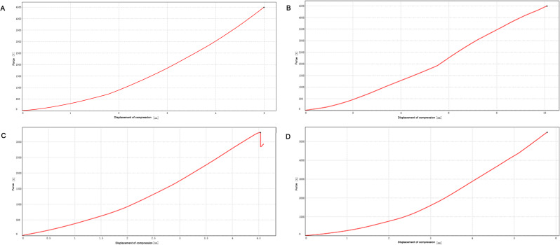
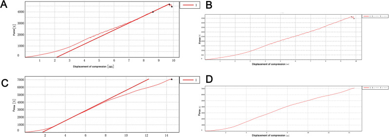
Comparing fixation methods for Sander II and III fractures, the force-displacement curve of the intramedullary nail group aligned more closely with standards. Under axial compression, bone stress was highest with the Sanders II locking plate and lowest with the intramedullary nail. Across models, the intramedullary nail consistently exhibited slightly higher stress than the locking plate. The intramedullary nailing group model of the overall stiffness was slightly greater than the locking plate. By comparing the compressive mechanical test performance of the two fixation methods, it was found that the plate fixation group had an abnormal load until 7092.895 N, which was 1.5 times more than the load of the intramedullary nail.
Both intramedullary fixation and locking plate fixation for Sanders II and III calcaneal fractures have certain biomechanical stability, and locking plate fixation has potential application value in clinical practice.
跟骨骨折固定在骨科领域仍然是一项具有挑战性的手术,计算工具越来越有助于优化术前规划。通过三维(3D)有限元分析比较髓内固定和锁定钢板固定治疗Sanders II型和III型跟骨骨折的生物力学稳定性,为临床应用提供理论依据。
使用Mimics软件(Materialise NV,比利时)对计算机断层扫描(CT)图像进行分割,基于阈值分割识别感兴趣区域。使用Mimics 10.01软件重建3D形态。随后,使用Geomagic2012软件(3D Systems,美国)去除噪声点、尖角和散点,获得平滑的表面图。该图以初始图形交换规范(IGES)格式保存,并导入Solidworks(达索系统公司,法国)进行模型组装和体积模型构建。建立Sanders II/III型跟骨骨折髓内固定和锁定钢板固定的三维有限元模型,并通过线性有限元分析进行分析。计算两种固定方法的强制位移、刚度和应力分布。此外,使用压缩力学实验对三维模型进行测试。
比较Sanders II型和III型骨折的固定方法,髓内钉组的力-位移曲线与标准更接近。在轴向压缩下,Sanders II型锁定钢板的骨应力最高,髓内钉的骨应力最低。在所有模型中,髓内钉的应力始终略高于锁定钢板。髓内钉固定组模型的整体刚度略大于锁定钢板。通过比较两种固定方法的压缩力学测试性能,发现钢板固定组在7092.895 N之前出现异常载荷,是髓内钉载荷的1.5倍。
髓内固定和锁定钢板固定治疗Sanders II型和III型跟骨骨折均具有一定的生物力学稳定性,锁定钢板固定在临床实践中具有潜在的应用价值。