Foliaki Simote T, Groveman Bradley R, Dews Emmett A, Williams Katie, El Soufi Hadil, Schwarz Benjamin, Leung Jacqueline M, Schneider Christine A, Schwartz Cindi L, Bohrnsen Eric, Kimzey Cole D, Race Brent, Haigh Cathryn L
Laboratory of Neurological Infections and Immunity, National Institute of Allergy and Infectious Diseases, Division of Intramural Research, Rocky Mountain Laboratories, National Institutes of Health, Hamilton, MT, USA.
Research Technologies Branch, National Institute of Allergy and Infectious Diseases, Division of Intramural Research, Rocky Mountain Laboratories, National Institutes of Health, Hamilton, MT, USA.
Acta Neuropathol Commun. 2024 Dec 20;12(1):192. doi: 10.1186/s40478-024-01905-w.
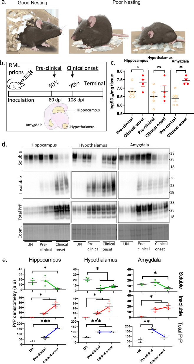
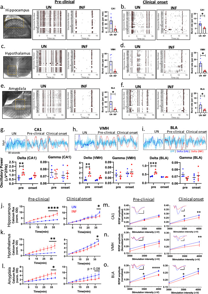
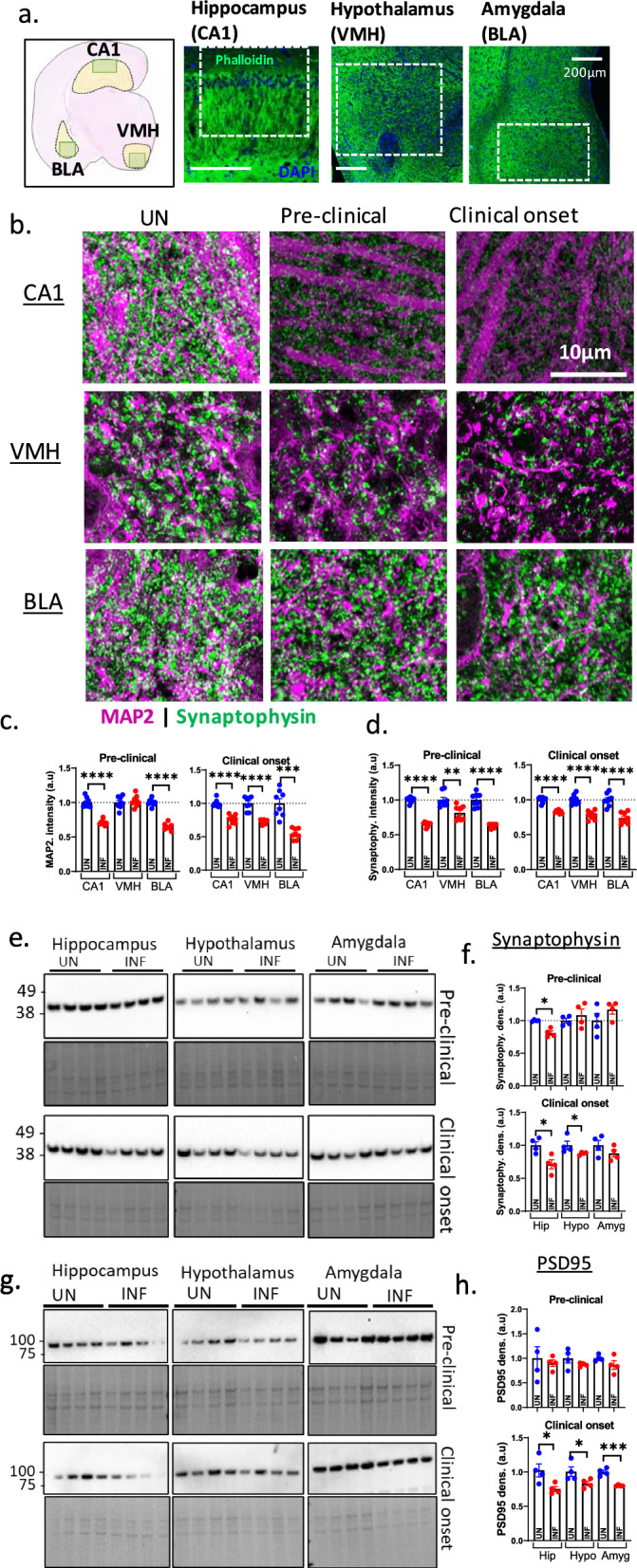
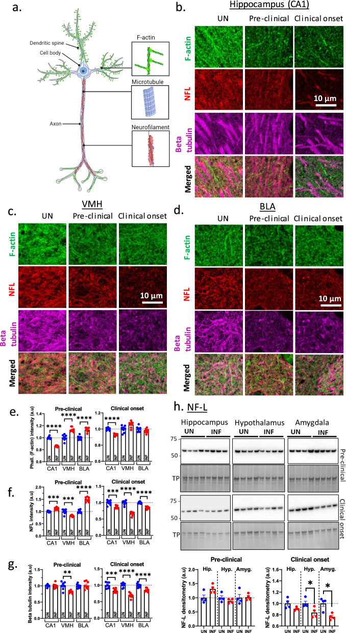
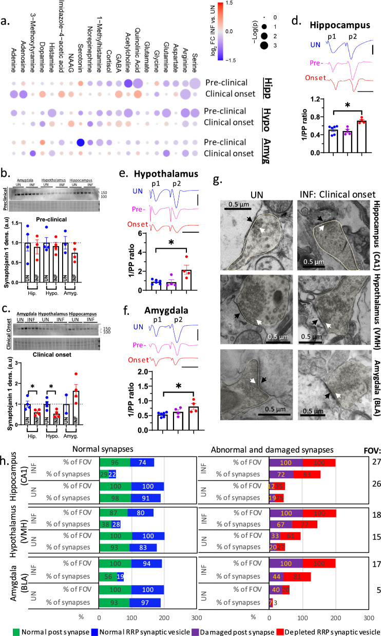
Misfolding of normal prion protein (PrP) to pathological isoforms (prions) causes prion diseases (PrDs) with clinical manifestations including cognitive decline and mood-related behavioral changes. Cognition and mood are linked to the neurophysiology of the limbic system. Little is known about how the disease affects the synaptic activity in brain parts associated with this system. We hypothesize that the dysfunction of synaptic transmission in the limbic regions correlates with the onset of reduced cognition and behavioral deficits. Here, we studied how prion infection in mice disrupts the synaptic function in three limbic regions, the hippocampus, hypothalamus, and amygdala, at a pre-clinical stage (mid-incubation period) and early clinical onset. PrD caused calcium flux dysregulation associated with lesser spontaneous synchronous neuronal firing and slowing neural oscillation at the pre-clinical stage in the hippocampal CA1, ventral medial hypothalamus, and basolateral amygdala (BLA). At clinical onset, synaptic transmission and synaptic plasticity became significantly disrupted. This correlated with a substantial depletion of the soluble prion protein, loss of total synapses, abnormal neurotransmitter levels and synaptic release, decline in synaptic vesicle recycling, and cytoskeletal damage. Further, the amygdala exhibited distinct disease-related changes in synaptic morphology and physiology compared with the other regions, but generally to a lesser degree, demonstrating how different rates of damage in the limbic system influence the evolution of clinical disease. Overall, PrD causes synaptic damage in three essential limbic regions starting at a preclinical stage and resulting in synaptic plasticity dysfunction correlated with early disease signs. Therapeutic drugs that alleviate these early neuronal dysfunctions may significantly delay clinical onset.
正常朊病毒蛋白(PrP)错误折叠为病理性异构体(朊病毒)会导致朊病毒疾病(PrDs),其临床表现包括认知能力下降和与情绪相关的行为变化。认知和情绪与边缘系统的神经生理学相关。关于该疾病如何影响与该系统相关的脑区突触活动,我们知之甚少。我们假设边缘区域突触传递功能障碍与认知能力下降和行为缺陷的发作相关。在此,我们研究了小鼠朊病毒感染如何在临床前阶段(潜伏期中期)和临床早期发作时破坏三个边缘区域(海马体、下丘脑和杏仁核)的突触功能。朊病毒疾病在临床前阶段,在海马体CA1区、腹内侧下丘脑和基底外侧杏仁核(BLA)导致钙通量失调,伴有较少的自发同步神经元放电和神经振荡减慢。在临床发作时,突触传递和突触可塑性受到显著破坏。这与可溶性朊病毒蛋白大量消耗、总突触丧失、神经递质水平和突触释放异常、突触小泡循环下降以及细胞骨架损伤相关。此外,与其他区域相比,杏仁核在突触形态和生理学上表现出明显的疾病相关变化,但总体程度较轻,这表明边缘系统不同的损伤速率如何影响临床疾病的发展。总体而言,朊病毒疾病从临床前阶段开始就在三个重要的边缘区域造成突触损伤,导致与早期疾病迹象相关的突触可塑性功能障碍。减轻这些早期神经元功能障碍的治疗药物可能会显著延迟临床发作。