Zhu Hao-Nan, Guo Yi-Fan, Lin YingMin, Sun Zhi-Chao, Zhu Xi, Li YuanZhe
The First School of Clinical Medicine, Zhejiang Chinese Medical University, Hangzhou, Zhejiang, China.
Department of Radiology, The First Affiliated Hospital of Zhejiang Chinese Medical University, (Zhejiang Provincial Hospital of Traditional Chinese Medicine), Hangzhou, Zhejiang, China.
J Bone Oncol. 2024 Nov 26;50:100653. doi: 10.1016/j.jbo.2024.100653. eCollection 2025 Feb.
Bone metastasis from breast cancer significantly elevates patient morbidity and mortality, making early detection crucial for improving outcomes. This study utilizes radiomics to analyze changes in the thoracic vertebral bone marrow microenvironment from chest computerized tomography (CT) images prior to bone metastasis in breast cancer, and constructs a model to predict metastasis.
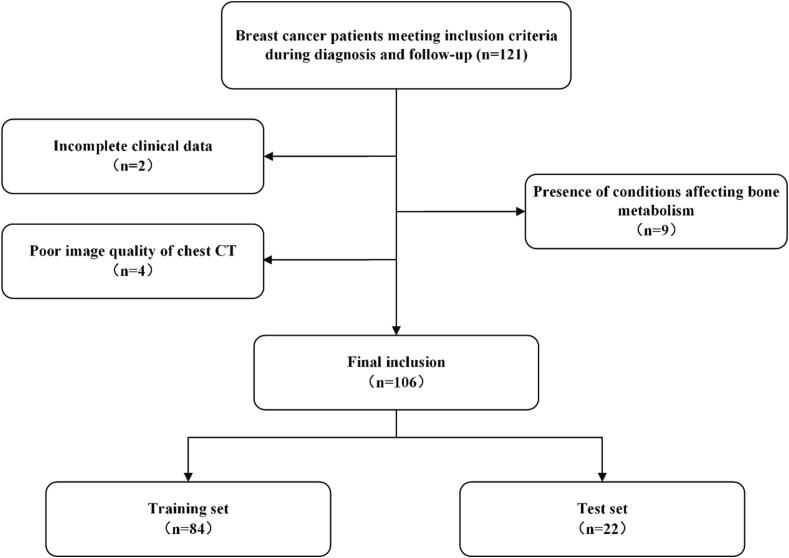
This study retrospectively gathered data from breast cancer patients who were diagnosed and continuously monitored for five years from January 2013 to September 2023. Radiomic features were extracted from the bone marrow of thoracic vertebrae on non-contrast chest CT scans. Multiple machine learning algorithms were utilized to construct various radiomics models for predicting the risk of bone metastasis, and the model with optimal performance was integrated with clinical features to develop a nomogram. The effectiveness of this combined model was assessed through receiver operating characteristic (ROC) analysis as well as decision curve analysis (DCA).
The study included a total of 106 patients diagnosed with breast cancer, among whom 37 developed bone metastases within five years. The radiomics model's area under the curve (AUC) for the test set, calculated using logistic regression, is 0.929, demonstrating superior predictive performance compared to alternative machine learning models. Furthermore, DCA demonstrated the potential of radiomics models in clinical application, with a greater clinical benefit in predicting bone metastasis than clinical model and nomogram.
CT-based radiomics can capture subtle changes in the thoracic vertebral bone marrow before breast cancer bone metastasis, offering a predictive tool for early detection of bone metastasis in breast cancer.
乳腺癌骨转移显著提高患者的发病率和死亡率,因此早期检测对于改善预后至关重要。本研究利用放射组学分析乳腺癌骨转移前胸部计算机断层扫描(CT)图像中胸椎骨髓微环境的变化,并构建预测转移的模型。
本研究回顾性收集了2013年1月至2023年9月期间被诊断并持续监测五年的乳腺癌患者的数据。从胸部非增强CT扫描的胸椎骨髓中提取放射组学特征。利用多种机器学习算法构建各种预测骨转移风险的放射组学模型,并将性能最佳的模型与临床特征相结合开发列线图。通过受试者操作特征(ROC)分析和决策曲线分析(DCA)评估该联合模型的有效性。
该研究共纳入106例确诊为乳腺癌的患者,其中37例在五年内发生骨转移。使用逻辑回归计算的测试集放射组学模型的曲线下面积(AUC)为0.929,与其他机器学习模型相比,显示出卓越的预测性能。此外,DCA证明了放射组学模型在临床应用中的潜力,在预测骨转移方面比临床模型和列线图具有更大的临床益处。
基于CT的放射组学可以捕捉乳腺癌骨转移前胸椎骨髓的细微变化,为乳腺癌骨转移的早期检测提供一种预测工具。