• 文献检索
  • 文档翻译
  • 深度研究
  • 学术资讯
  • Suppr Zotero 插件Zotero 插件
  • 邀请有礼
  • 套餐&价格
  • 历史记录
应用&插件
Suppr Zotero 插件Zotero 插件浏览器插件Mac 客户端Windows 客户端微信小程序
定价
高级版会员购买积分包购买API积分包
服务
文献检索文档翻译深度研究API 文档MCP 服务
关于我们
关于 Suppr公司介绍联系我们用户协议隐私条款
关注我们

Suppr 超能文献

核心技术专利:CN118964589B侵权必究
粤ICP备2023148730 号-1Suppr @ 2026

文献检索

告别复杂PubMed语法,用中文像聊天一样搜索,搜遍4000万医学文献。AI智能推荐,让科研检索更轻松。

立即免费搜索

文件翻译

保留排版,准确专业,支持PDF/Word/PPT等文件格式,支持 12+语言互译。

免费翻译文档

深度研究

AI帮你快速写综述,25分钟生成高质量综述,智能提取关键信息,辅助科研写作。

立即免费体验

用于临床环境中智能手机细胞浓度分析的无泵微流控技术(SmartFlow):设计、开发与评估

Pump-Free Microfluidics for Cell Concentration Analysis on Smartphones in Clinical Settings (SmartFlow): Design, Development, and Evaluation.

作者信息

Wu Sixuan, Song Kefan, Cobb Jason, Adams Alexander T

机构信息

School of Interactive Computing, Georgia Institute of Technology, Atlanta, GA, United States.

Wallace H Coulter Department of Biomedical Engineering, Georgia Institute of Technology, Atlanta, GA, United States.

出版信息

JMIR Biomed Eng. 2024 Dec 23;9:e62770. doi: 10.2196/62770.

DOI:10.2196/62770
PMID:39715548
原文链接:https://pmc.ncbi.nlm.nih.gov/articles/PMC11704648/
Abstract

BACKGROUND

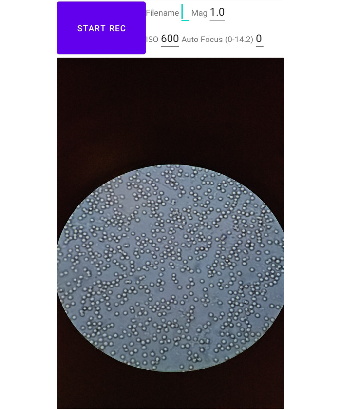
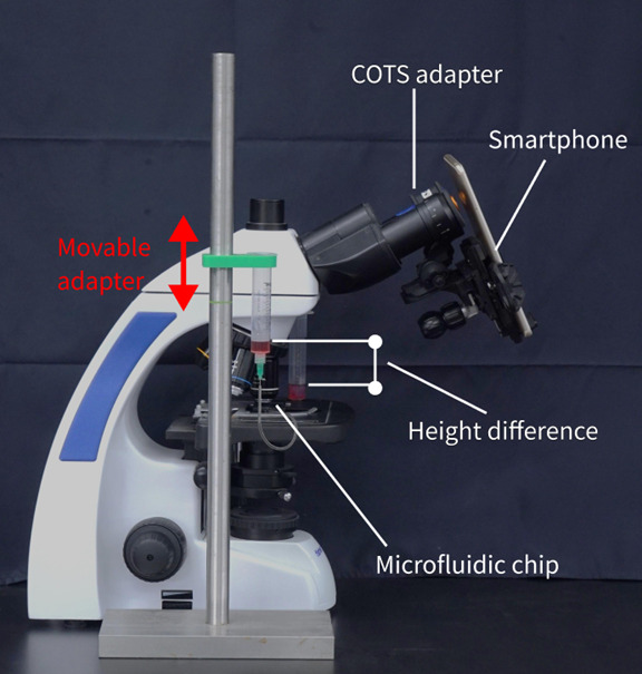
Cell concentration in body fluid is an important factor for clinical diagnosis. The traditional method involves clinicians manually counting cells under microscopes, which is labor-intensive. Automated cell concentration estimation can be achieved using flow cytometers; however, their high cost limits accessibility. Microfluidic systems, although cheaper than flow cytometers, still require high-speed cameras and syringe pumps to drive the flow and ensure video quality. In this paper, we present SmartFlow, a low-cost solution for cell concentration estimation using smartphone-based computer vision on 3D-printed, pump-free microfluidic platforms.

OBJECTIVE

The objective was to design and fabricate microfluidic chips, coupled with clinical utilities, for cell counting and concentration analysis. We answered the following research questions (RQs): RQ1, Can gravity drive the flow within the microfluidic chips, eliminating the need for external pumps? RQ2, How does the microfluidic chip design impact video quality for cell analysis? RQ3, Can smartphone-captured videos be used to estimate cell count and concentration in microfluidic chips?

METHODS

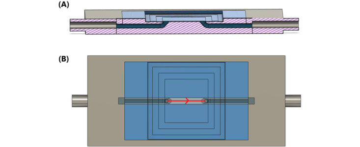
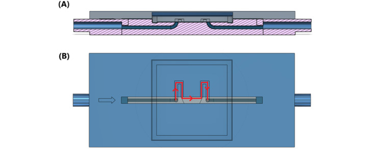
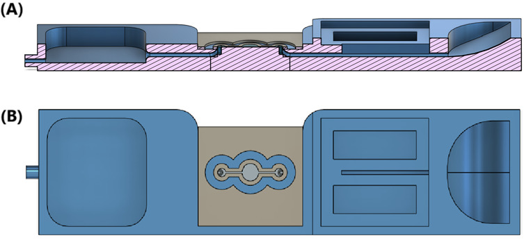
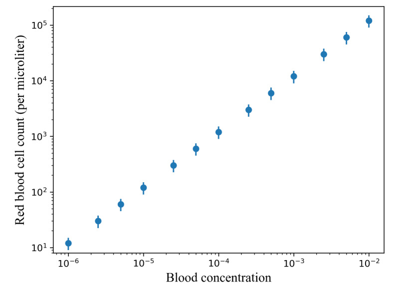
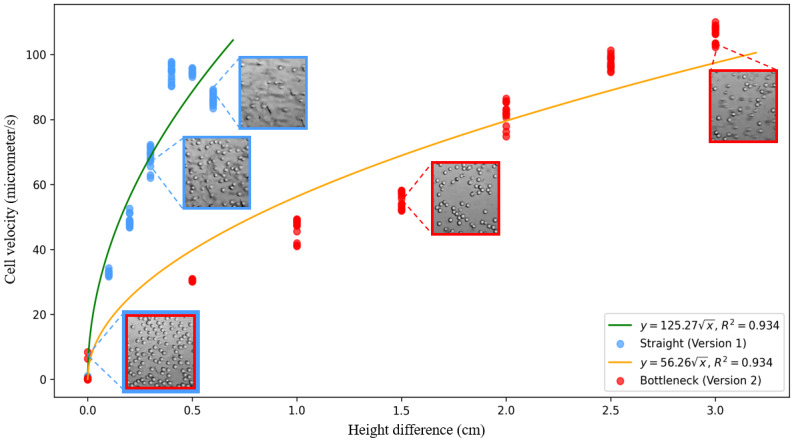
To answer the 3 RQs, 2 experiments were conducted. In the cell flow velocity experiment, diluted sheep blood flowed through the microfluidic chips with and without a bottleneck design to answer RQ1 and RQ2, respectively. In the cell concentration analysis experiment, sheep blood diluted into 13 concentrations flowed through the microfluidic chips while videos were recorded by smartphones for the concentration measurement.

RESULTS

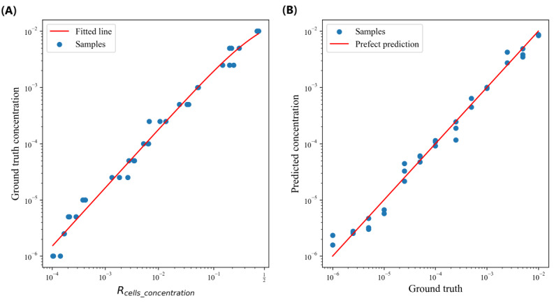
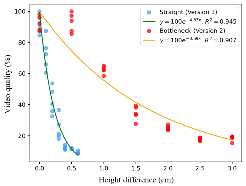
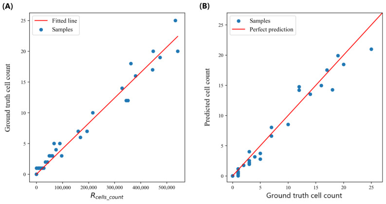
In the cell flow velocity experiment, we designed and fabricated 2 versions of microfluidic chips. The ANOVA test (Straight: F=6144.45, P<.001; Bottleneck: F=3475.78, P<.001) showed the height difference had a significant impact on the cell velocity, which implied gravity could drive the flow. The video sharpness analysis demonstrated that video quality followed an exponential decay with increasing height differences (video quality=100e) and a bottleneck design could effectively preserve video quality (Straight: R=0.95, k=4.33; Bottleneck: R=0.91, k=0.59). Samples from the 13 cell concentrations were used for cell counting and cell concentration estimation analysis. The accuracy of cell counting (n=35, 60-second samples, R=0.96, mean absolute error=1.10, mean squared error=2.24, root mean squared error=1.50) and cell concentration regression (n=39, 150-second samples, R=0.99, mean absolute error=0.24, mean squared error=0.11, root mean squared error=0.33 on a logarithmic scale, mean average percentage error=0.25) were evaluated using 5-fold cross-validation by comparing the algorithmic estimation to ground truth.

CONCLUSIONS

In conclusion, we demonstrated the importance of the flow velocity in a microfluidic system, and we proposed SmartFlow, a low-cost system for computer vision-based cellular analysis. The proposed system could count the cells and estimate cell concentrations in the samples.

摘要

背景

体液中的细胞浓度是临床诊断的一个重要因素。传统方法是临床医生在显微镜下手动计数细胞,这一过程 labor-intensive。使用流式细胞仪可以实现细胞浓度的自动估计;然而,其高昂的成本限制了其普及。微流控系统虽然比流式细胞仪便宜,但仍需要高速相机和注射泵来驱动流体并确保视频质量。在本文中,我们展示了 SmartFlow,这是一种基于智能手机计算机视觉的低成本解决方案,用于在 3D 打印的无泵微流控平台上估计细胞浓度。

目的

目的是设计并制造与临床应用相结合的微流控芯片,用于细胞计数和浓度分析。我们回答了以下研究问题(RQs):问题 1,重力能否驱动微流控芯片内的流体流动,从而无需外部泵?问题 2,微流控芯片设计如何影响细胞分析的视频质量?问题 3,智能手机拍摄的视频能否用于估计微流控芯片中的细胞计数和浓度?

方法

为回答这 3 个问题,进行了 2 项实验。在细胞流速实验中,稀释的羊血分别流过有无瓶颈设计的微流控芯片,以分别回答问题 1 和问题 2。在细胞浓度分析实验中,稀释成 13 种浓度的羊血流过微流控芯片,同时智能手机记录视频以进行浓度测量。

结果

在细胞流速实验中,我们设计并制造了 2 个版本的微流控芯片。方差分析测试(无瓶颈:F = 6144.45,P <.001;有瓶颈:F = 3475.78,P <.001)表明高度差对细胞流速有显著影响,这意味着重力可以驱动流体流动。视频清晰度分析表明,视频质量随高度差增加呈指数衰减(视频质量 = 100e),并且瓶颈设计可以有效保持视频质量(无瓶颈:R = 0.95,k = 4.33;有瓶颈:R = 0.91,k = 0.59)。来自 13 种细胞浓度的样本用于细胞计数和细胞浓度估计分析。通过将算法估计与真实值进行比较,使用 5 折交叉验证评估细胞计数的准确性(n = 35,60 秒样本,R = 0.96,平均绝对误差 = 1.10,均方误差 = 2.24,均方根误差 = 1.50)和细胞浓度回归(n = 39,150 秒样本,R = 0.99,平均绝对误差 = 0.24,均方误差 = 0.11,对数尺度下的均方根误差 = 0.33,平均平均百分比误差 = 0.25)。

结论

总之,我们证明了微流控系统中流速的重要性,并提出了 SmartFlow,这是一种用于基于计算机视觉的细胞分析的低成本系统。所提出的系统可以对样本中的细胞进行计数并估计细胞浓度。

https://cdn.ncbi.nlm.nih.gov/pmc/blobs/a568/11704648/6b2fde7f568b/biomedeng_v9i1e62770_fig12.jpg
https://cdn.ncbi.nlm.nih.gov/pmc/blobs/a568/11704648/3f312b4b6913/biomedeng_v9i1e62770_fig1.jpg
https://cdn.ncbi.nlm.nih.gov/pmc/blobs/a568/11704648/a5d024846bf4/biomedeng_v9i1e62770_fig2.jpg
https://cdn.ncbi.nlm.nih.gov/pmc/blobs/a568/11704648/e7e2c91185c3/biomedeng_v9i1e62770_fig3.jpg
https://cdn.ncbi.nlm.nih.gov/pmc/blobs/a568/11704648/6aecbd9e846f/biomedeng_v9i1e62770_fig4.jpg
https://cdn.ncbi.nlm.nih.gov/pmc/blobs/a568/11704648/11367996916b/biomedeng_v9i1e62770_fig5.jpg
https://cdn.ncbi.nlm.nih.gov/pmc/blobs/a568/11704648/85a96dd01163/biomedeng_v9i1e62770_fig6.jpg
https://cdn.ncbi.nlm.nih.gov/pmc/blobs/a568/11704648/0c5127b44f35/biomedeng_v9i1e62770_fig7.jpg
https://cdn.ncbi.nlm.nih.gov/pmc/blobs/a568/11704648/f4c51dd1925b/biomedeng_v9i1e62770_fig8.jpg
https://cdn.ncbi.nlm.nih.gov/pmc/blobs/a568/11704648/8d2109614dac/biomedeng_v9i1e62770_fig9.jpg
https://cdn.ncbi.nlm.nih.gov/pmc/blobs/a568/11704648/9c336b83b2c4/biomedeng_v9i1e62770_fig10.jpg
https://cdn.ncbi.nlm.nih.gov/pmc/blobs/a568/11704648/ee8eed05ff5b/biomedeng_v9i1e62770_fig11.jpg
https://cdn.ncbi.nlm.nih.gov/pmc/blobs/a568/11704648/6b2fde7f568b/biomedeng_v9i1e62770_fig12.jpg

相似文献

1
Pump-Free Microfluidics for Cell Concentration Analysis on Smartphones in Clinical Settings (SmartFlow): Design, Development, and Evaluation.用于临床环境中智能手机细胞浓度分析的无泵微流控技术(SmartFlow):设计、开发与评估
JMIR Biomed Eng. 2024 Dec 23;9:e62770. doi: 10.2196/62770.
2
Equity-Driven Sensing System for Measuring Skin Tone-Calibrated Peripheral Blood Oxygen Saturation (OptoBeat): Development, Design, and Evaluation Study.用于测量肤色校准外周血氧饱和度的公平驱动传感系统(OptoBeat):开发、设计与评估研究
JMIR Biomed Eng. 2022 Apr 22;7(1):e34934. doi: 10.2196/34934.
3
Digital monitoring of the microchannel filling flow dynamics using a non-contactless smartphone-based nano-liter precision flow velocity meter.使用非接触式基于智能手机的纳升级精密流速计对微通道填充流动动力学进行数字监测。
Biosens Bioelectron. 2024 May 15;252:116130. doi: 10.1016/j.bios.2024.116130. Epub 2024 Feb 13.
4
Open-source spring-driven syringe pump with 3D-printed components for microfluidic applications.用于微流体应用的具有3D打印部件的开源弹簧驱动注射器泵。
HardwareX. 2024 Jul 6;19:e00550. doi: 10.1016/j.ohx.2024.e00550. eCollection 2024 Sep.
5
Folic acid supplementation and malaria susceptibility and severity among people taking antifolate antimalarial drugs in endemic areas.在流行地区,服用抗叶酸抗疟药物的人群中,叶酸补充剂与疟疾易感性和严重程度的关系。
Cochrane Database Syst Rev. 2022 Feb 1;2(2022):CD014217. doi: 10.1002/14651858.CD014217.
6
[Standard technical specifications for methacholine chloride (Methacholine) bronchial challenge test (2023)].[氯化乙酰甲胆碱支气管激发试验标准技术规范(2023年)]
Zhonghua Jie He He Hu Xi Za Zhi. 2024 Feb 12;47(2):101-119. doi: 10.3760/cma.j.cn112147-20231019-00247.
7
Smartphone based on-chip fluorescence imaging and capillary flow velocity measurement for detecting ROR1+ cancer cells from buffy coat blood samples on dual-layer paper microfluidic chip.基于智能手机的片上荧光成像和毛细管流动速度测量技术,用于在双层纸微流控芯片上从缓冲血样中检测 ROR1+癌细胞。
Biosens Bioelectron. 2020 Apr 1;153:112042. doi: 10.1016/j.bios.2020.112042. Epub 2020 Jan 22.
8
[Applications of microfluidic paper-based chips in environmental analysis and detection].基于微流控纸芯片在环境分析与检测中的应用
Se Pu. 2021 Aug;39(8):802-815. doi: 10.3724/SP.J.1123.2020.09004.
9
Compact and automated particle counting platform using smartphone-microscopy.采用智能手机显微镜的紧凑型自动化颗粒计数平台。
Talanta. 2021 Jun 1;228:122244. doi: 10.1016/j.talanta.2021.122244. Epub 2021 Feb 26.
10
Cell-sorting centrifugal microfluidic chip with a flow rectifier.带有整流器的细胞分选离心微流控芯片
Lab Chip. 2021 Jun 1;21(11):2129-2141. doi: 10.1039/d1lc00217a.

本文引用的文献

1
Interfacial tension driven open droplet microfluidics.界面张力驱动的开放液滴微流控技术。
Adv Mater Interfaces. 2023 Mar 6;10(7). doi: 10.1002/admi.202202234. Epub 2023 Jan 22.
2
Computer vision meets microfluidics: a label-free method for high-throughput cell analysis.计算机视觉与微流控技术相结合:一种用于高通量细胞分析的无标记方法。
Microsyst Nanoeng. 2023 Sep 21;9:116. doi: 10.1038/s41378-023-00562-8. eCollection 2023.
3
Smartphone-based platforms implementing microfluidic detection with image-based artificial intelligence.
基于智能手机的平台,结合基于图像的人工智能,实现微流控检测。
Nat Commun. 2023 Mar 11;14(1):1341. doi: 10.1038/s41467-023-36017-x.
4
Gravity-driven microfluidic device placed on a slow-tilting table enables constant unidirectional perfusion culture of human induced pluripotent stem cells.放置在缓慢倾斜平台上的重力驱动微流控装置能够实现人诱导多能干细胞的恒定单向灌注培养。
J Biosci Bioeng. 2023 Feb;135(2):151-159. doi: 10.1016/j.jbiosc.2022.11.007. Epub 2022 Dec 29.
5
Novel Pumping Methods for Microfluidic Devices: A Comprehensive Review.新型微流控芯片泵送方法:全面综述
Biosensors (Basel). 2022 Nov 1;12(11):956. doi: 10.3390/bios12110956.
6
Deep learning detector for high precision monitoring of cell encapsulation statistics in microfluidic droplets.深度学习检测器,用于高精度监测微流控液滴中的细胞封装统计数据。
Lab Chip. 2022 Oct 25;22(21):4067-4080. doi: 10.1039/d2lc00462c.
7
Microfluidic paper-based aptasensor devices for multiplexed detection of pathogenic bacteria.用于多重检测病原菌的基于微流控纸的适配体传感器装置
Biosens Bioelectron. 2022 Jul 1;207:114214. doi: 10.1016/j.bios.2022.114214. Epub 2022 Mar 24.
8
Direct capture and smartphone quantification of airborne SARS-CoV-2 on a paper microfluidic chip.直接捕获并通过智能手机对纸基微流控芯片上的空气传播 SARS-CoV-2 进行定量检测。
Biosens Bioelectron. 2022 Mar 15;200:113912. doi: 10.1016/j.bios.2021.113912. Epub 2021 Dec 24.
9
Gravity-Driven Microfluidic Siphons: Fluidic Characterization and Application to Quantitative Immunoassays.重力驱动微流控虹吸管:流体特性分析及其在定量免疫分析中的应用。
ACS Sens. 2021 Dec 24;6(12):4338-4348. doi: 10.1021/acssensors.1c01524. Epub 2021 Dec 2.
10
Label-free classification of dead and live colonic adenocarcinoma cells based on 2D light scattering and deep learning analysis.基于二维光散射和深度学习分析的结肠腺癌死活细胞无标记分类。
Cytometry A. 2021 Nov;99(11):1134-1142. doi: 10.1002/cyto.a.24475. Epub 2021 Jun 19.