Department of Biomedical Engineering, The University of Arizona, Tucson, AZ, 85721, United States.
Hematology Oncology Division, Mayo Clinic, Phoenix, AZ, 85054, United States.
Biosens Bioelectron. 2020 Apr 1;153:112042. doi: 10.1016/j.bios.2020.112042. Epub 2020 Jan 22.
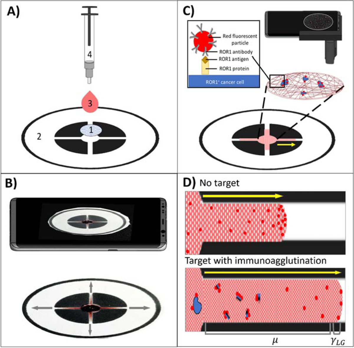
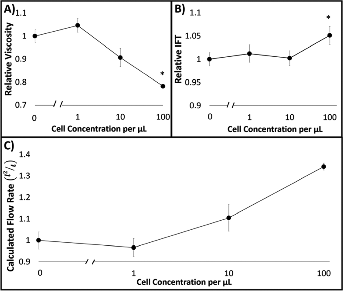
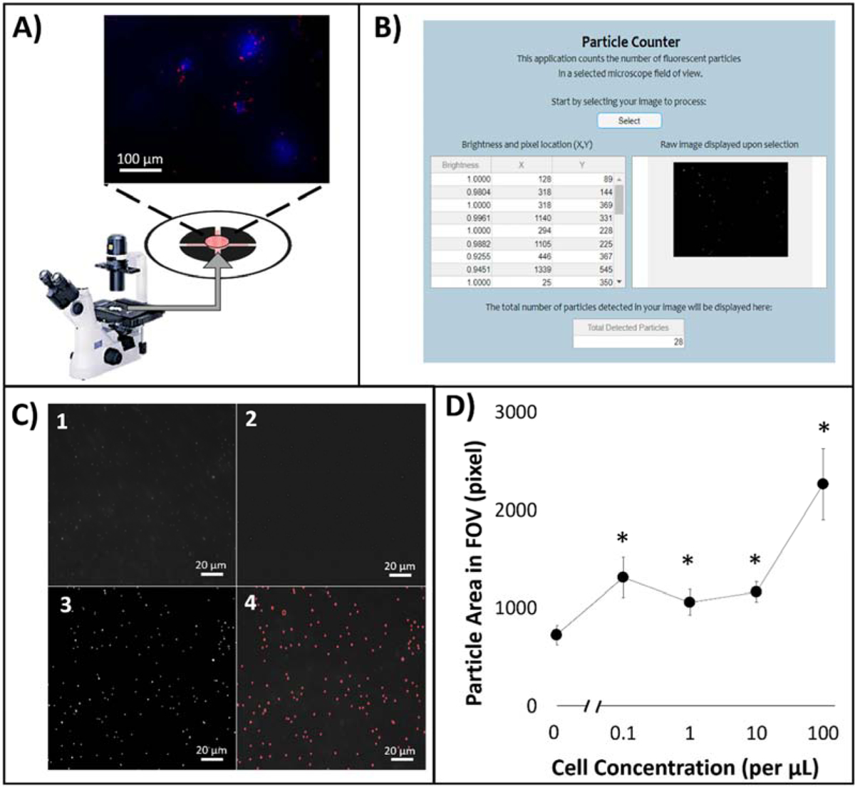
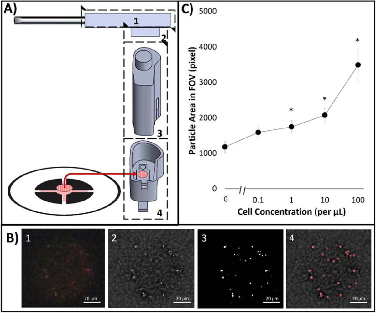
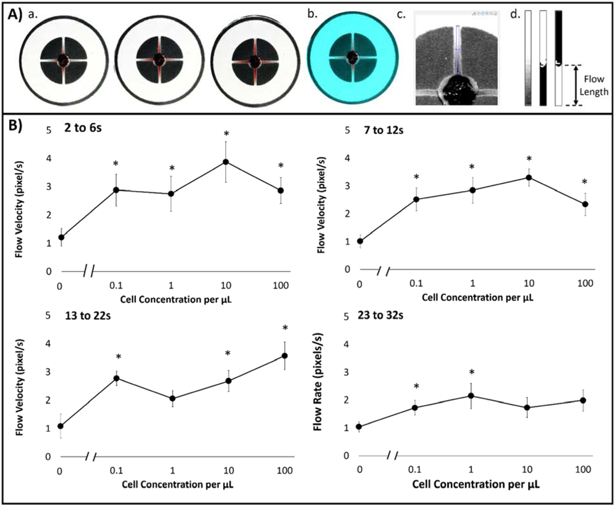
Diagnosis of hematological cancer requires complete white blood cell count, followed by flow cytometry with multiple markers, and cytology. It requires substantial time and specialized training. A dual-layer paper microfluidic chip was developed as a quicker, low-cost, and field-deployable alternative to detect ROR1+ (receptor tyrosine-like orphan receptor one) cancer cells from the undiluted and untreated buffy coat blood samples. The first capture layer consisted of a GF/D glass fiber substrate, preloaded with cancer specific anti-ROR1 conjugated fluorescent particles to its center for cancer cell capture and direct smartphone fluorescence imaging. The second flow layer was comprised of a grade 1 cellulose chromatography paper with wax-printed four channels for wicking and capillary flow-based detection. The flow velocity was used as measure of antigen concentration in the buffy coat sample. In this manner, intact cells and their antigens were separated and independently analyzed by both imaging and flow velocity analyses. A custom-made smartphone-based fluorescence microscope and automated image processing and particle counter software were developed to enumerate particles on paper, with the limit of detection of 1 cell/μL. Flow velocity analysis showed even greater sensitivity, with the limit of detection of 0.1 cells/μL in the first 6 s of assay. Comparison with capillary flow model revealed great alignment with experimental data and greater correlation to viscosity than interfacial tension. Our proposed device is able to capture and on-chip image ROR1+ cancer cells within a complex sample matrix (buffy coat) while simultaneously quantifying cell concentration in a point-of-care manner.
血液癌症的诊断需要进行完整的白细胞计数,然后进行带有多个标志物的流式细胞术和细胞学检查。这需要大量的时间和专门的培训。本研究开发了一种双层纸质微流控芯片,作为一种更快、低成本、可现场部署的替代方案,用于从未经稀释和未经处理的血涂片样本中检测 ROR1+(受体酪氨酸样孤儿受体 1)癌细胞。第一层捕获层由 GF/D 玻璃纤维基质组成,预先加载有针对 ROR1 的癌症特异性荧光偶联颗粒,位于中心位置,用于捕获癌细胞并直接通过智能手机荧光成像进行检测。第二层流层由一级纤维素层析纸组成,纸上印有四个蜡印通道,用于虹吸和基于毛细流动的检测。流动速度用作缓冲层样本中抗原浓度的测量值。通过这种方式,可以对完整细胞及其抗原进行分离,并通过成像和流动速度分析进行独立分析。本研究开发了一种定制的基于智能手机的荧光显微镜以及自动化图像处理和粒子计数器软件,用于对纸上的粒子进行计数,检测限为 1 个细胞/μL。流动速度分析显示出更高的灵敏度,在分析的前 6 秒内,检测限为 0.1 个细胞/μL。与毛细管流动模型的比较表明,与实验数据非常吻合,与界面张力相比,与粘度的相关性更高。本研究提出的设备能够在复杂的样本基质(血涂片)中捕获和芯片上成像 ROR1+癌细胞,同时以即时护理的方式定量细胞浓度。