Hao Qi, Zhang Yan, Li Xingpeng, Sun Xiaoli, Hong Nan, Wang Rengui
Department of Radiology, Peking University People's Hospital, No.11 Xizhimen South Street, Xicheng District, Beijing, 100044, China.
Department of Radiology, Qilu Hospital of Shandong University, Jinan, China.
BMC Med Imaging. 2024 Dec 23;24(1):348. doi: 10.1186/s12880-024-01504-0.
To investigate the diagnostic value of CT lymphangiography (CTL) and non-contrast MR lymphangiography (MRL) in lymphatic plastic bronchitis.
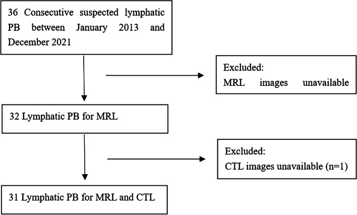
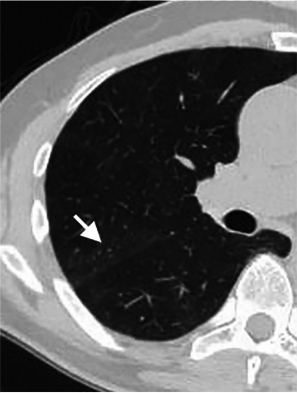
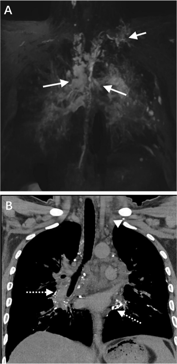
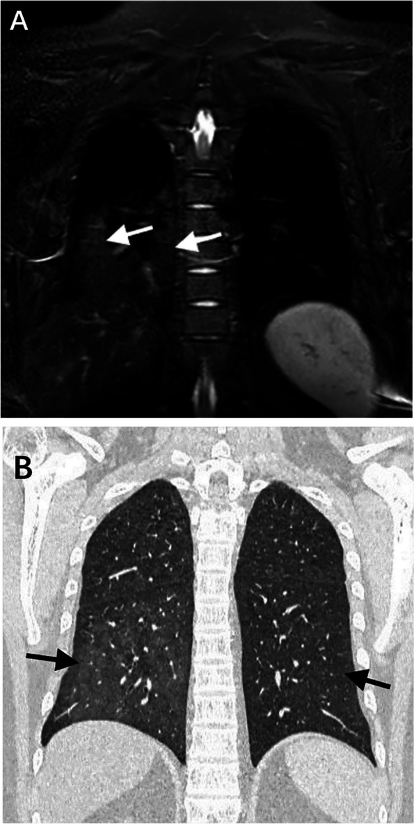
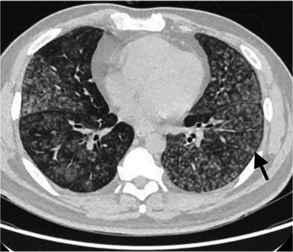
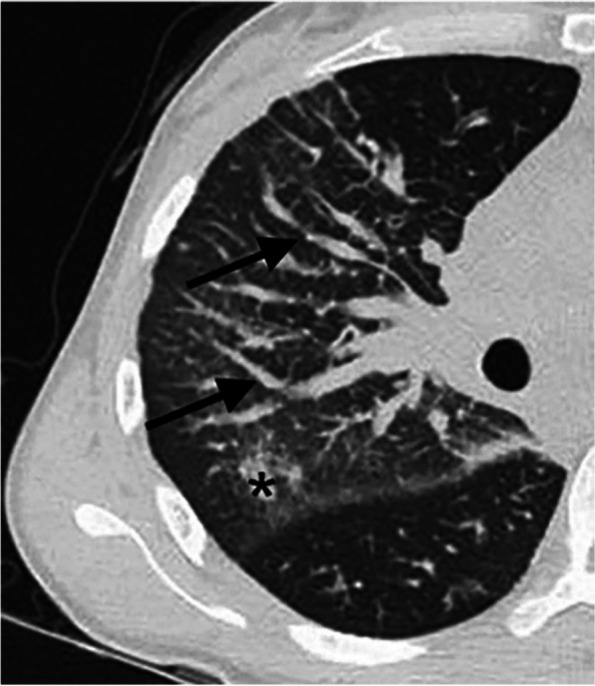
The clinical and imaging data of 31 patients with lymphatic plastic bronchitis diagnosed by clinical, imaging and pathological results were retrospectively analyzed. All patients underwent CTL and MRL. The imaging findings of patients include: (i) abnormal lymphatic reflux of the bronchial mediastinal trunk, the subclavian trunk, the cervical trunk, the thoracic duct and the right lymphatic duct; Abnormal CTL reflux refers to abnormal iodide deposition outside the normal lymphatic reflux pathway; If the MRL can observe abnormal lymphatic dilatation, hyperplasia, or morphological abnormalities, it is assumed that abnormal lymphatic reflux may be present.; (ii) abnormal morphological changes of lymphatic vessels at the extremity of the thoracic duct, the extremity of the right lymphatic duct and the mediastinum, such as spot-like or tubular, cystic changes; (iii) abnormal CTL and MRL signs in the lungs. The Mcnemar test was used to compare the parameters between CTL and MRL. P< 0.05 was statistically significant. The Kappa test was used to evaluate the consistency of CTL and MRL in evaluating lymphatic plastic bronchitis.
MRL was superior to CTL in detecting abnormal lymphatic reflux in the right lymphatic vessel, thoracic duct, cervical trunk and subclavian trunk (P< 0.05).and the diagnostic consistency was general (Kappa < 0.40). There was no significant difference between MRL and CTL in the detection of abnormal lymphatic reflux in the bronchial mediastinal trunk (P> 0.05), and the diagnostic consistency was good (Kappa > 0.60). MRL was superior to CTL in detecting lymphatic abnormalities such as cystic changes at the extremity of the thoracic duct, spot-like or tubular changes at the extremity of the right lymphatic duct, cystic changes at the extremity of the right lymphatic duct, and cystic changes in the mediastinum (P< 0.05), and the diagnostic consistency was poor, fair, fair, and moderate (Kappa < 0.60), respectively. MRL and CTL showed abnormal signs in the lung: CTL was superior to MRL in showing the thickening of interlobular septum, lung nodules and airway stenosis (P< 0.05), and the diagnostic consistency was moderate, moderate and poor (Kappa < 0.60). There was no significant difference between CTL and MRL in atelectasis, consolidation in lobar and segmental distribution, consolidation in non-lobar and segmental distribution, and the thickening of the bronchovascular bundle (P> 0.05), and the diagnostic consistency was very good, very good, good, good (Kappa > 0.60). There was no significant difference between CTL and MRL in ground glass opacity, airway wall thickening and intralobular interstitial thickening (P> 0.05), and the diagnostic consistency was average, fair and poor (Kappa < 0.40).
The MRL is superior to CTL in showing the abnormalities of the thoracic duct, the right lymphatic duct and other abnormal lymphatic vessels. CTL is superior to MRL in the detection of pulmonary abnormalities. The combination of CTL and MRL can provide more comprehensive imaging information for diagnosing and treating lymphatic plastic bronchitis.
探讨CT淋巴管造影(CTL)及非增强磁共振淋巴管造影(MRL)在淋巴塑型支气管炎中的诊断价值。
回顾性分析31例经临床、影像及病理结果确诊为淋巴塑型支气管炎患者的临床及影像资料。所有患者均接受了CTL及MRL检查。患者的影像表现包括:(i)支气管纵隔干、锁骨下干、颈干、胸导管及右淋巴管的异常淋巴回流;CTL异常回流是指在正常淋巴回流途径外出现异常碘沉积;若MRL能观察到异常淋巴管扩张、增生或形态异常,则推测可能存在异常淋巴回流;(ii)胸导管末端、右淋巴管末端及纵隔处淋巴管的形态异常改变,如斑点状或管状、囊性改变;(iii)肺部的CTL及MRL异常征象。采用McNemar检验比较CTL与MRL之间的参数。P<0.05具有统计学意义。采用Kappa检验评估CTL与MRL在评估淋巴塑型支气管炎方面的一致性。
MRL在检测右淋巴管、胸导管、颈干及锁骨下干的异常淋巴回流方面优于CTL(P<0.05),诊断一致性一般(Kappa<0.40)。MRL与CTL在检测支气管纵隔干的异常淋巴回流方面无显著差异(P>0.05),诊断一致性良好(Kappa>0.60)。MRL在检测胸导管末端的囊性改变、右淋巴管末端的斑点状或管状改变、右淋巴管末端的囊性改变及纵隔的囊性改变等淋巴异常方面优于CTL(P<0.05),诊断一致性分别为差、一般、一般及中等(Kappa<0.60)。MRL和CTL在肺部均显示有异常征象:CTL在显示小叶间隔增厚、肺结节及气道狭窄方面优于MRL(P<0.05),诊断一致性分别为中等、中等及差(Kappa<0.60)。CTL与MRL在肺不张、肺叶及肺段分布的实变、非肺叶及肺段分布的实变以及支气管血管束增粗方面无显著差异(P>0.05),诊断一致性分别为非常好、非常好、良好、良好(Kappa>0.60)。CTL与MRL在磨玻璃影、气道壁增厚及小叶内间质增厚方面无显著差异(P>0.05),诊断一致性分别为一般、一般及差(Kappa<0.40)。
MRL在显示胸导管、右淋巴管及其他异常淋巴管的异常方面优于CTL。CTL在检测肺部异常方面优于MRL。CTL与MRL联合应用可为淋巴塑型支气管炎的诊断及治疗提供更全面的影像信息。