Miyamae Nao, Ogai Kazuhiro, Kunimitsu Mao, Fujiwara Masayuki, Nagai Makoto, Okamoto Shigefumi, Okuwa Mayumi, Oe Makoto
Graduate School of Medical Sciences, Kanazawa University, Kanazawa, Japan.
Department of Fundamental Nursing, School of Nursing, Hyogo Medical University, Kobe, Japan.
Asia Pac J Oncol Nurs. 2024 Nov 19;12:100625. doi: 10.1016/j.apjon.2024.100625. eCollection 2025 Dec.
Severe radiodermatitis with erosion is a painful condition that affects quality of life; therefore, developing methods for its prevention is an urgent issue. Therefore, this study aimed to determine the morphological characteristics of the development and healing processes of severe radiodermatitis in patients with head and neck cancer and to explore the association between skin barrier function and development of severe radiodermatitis.
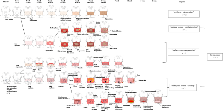
In this prospective observational study, the cervical regions of patients with head and neck cancer who underwent radiotherapy at a university hospital from October 2022 to March 2023 were photographed, and morphological characteristics of the development and healing process of severe radiodermatitis were extracted using the qualitative sketch method. Skin barrier function, including skin microbiota and dermal echogenicity, was investigated before initiating radiotherapy, and its relationship with radiodermatitis was examined using the Mann-Whitney test or Fisher's exact probability test.
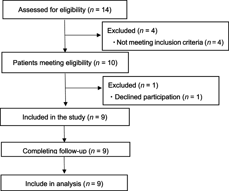
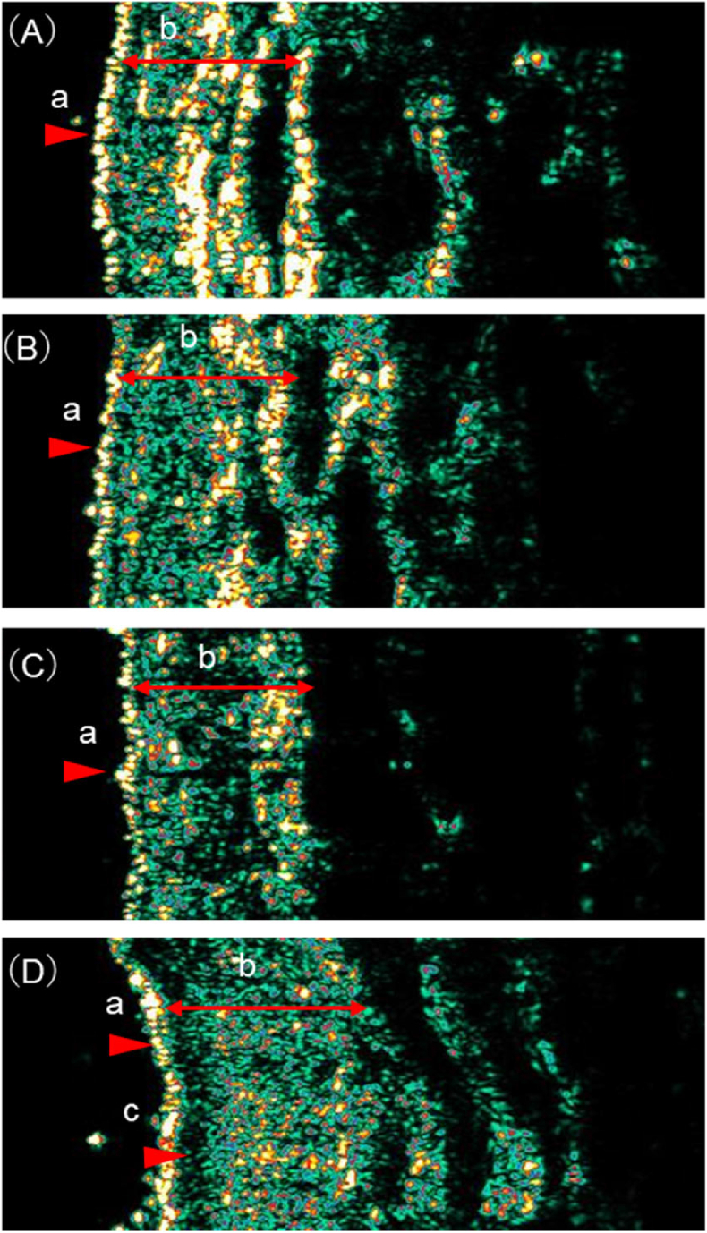
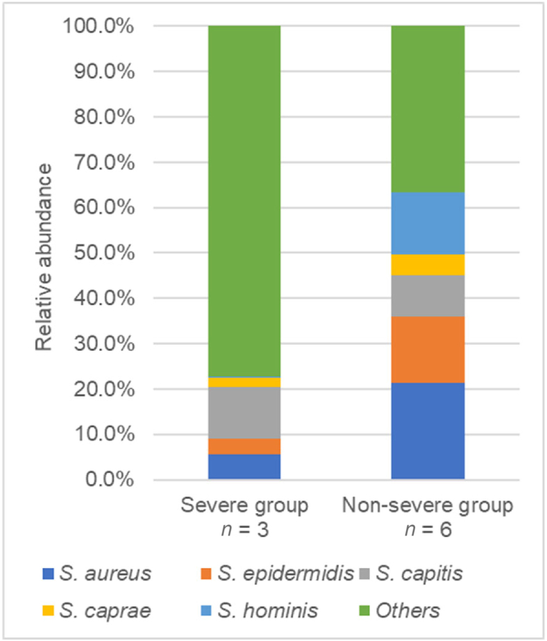
Nine patients were followed for a median of 61 (range 55-87) days with a total of 88 observations. The morphological characteristics of severe radiodermatitis were "localized erosion-epithelialization" and "widespread erosion-crusting," and compared to non-severe radiodermatitis, with low levels of ( = 0.024), ( = 0.024), and reduced dermal echogenicity ( = 0.036). Furthermore, the "widespread erosion-crusting" was associated with a subepidermal low echogenic band.
To prevent severe radiodermatitis, in addition to moisturizing the irradiated area and protecting it from mechanical irritation, improving skin barrier function before radiotherapy initiation may be effective.
伴有糜烂的严重放射性皮炎是一种影响生活质量的疼痛性病症;因此,开发其预防方法是一个紧迫的问题。因此,本研究旨在确定头颈癌患者严重放射性皮炎发生和愈合过程的形态学特征,并探讨皮肤屏障功能与严重放射性皮炎发生之间的关联。
在这项前瞻性观察研究中,对2022年10月至2023年3月在某大学医院接受放疗的头颈癌患者的颈部区域进行拍照,并使用定性素描法提取严重放射性皮炎发生和愈合过程的形态学特征。在放疗开始前调查包括皮肤微生物群和真皮回声性在内的皮肤屏障功能,并使用Mann-Whitney检验或Fisher精确概率检验检查其与放射性皮炎的关系。
9例患者接受了中位61天(范围55 - 87天)的随访,共进行了88次观察。严重放射性皮炎的形态学特征为“局限性糜烂 - 上皮形成”和“广泛糜烂 - 结痂”,与非严重放射性皮炎相比,其 ( = 0.024)、 ( = 0.024)水平较低,真皮回声性降低( = 0.036)。此外,“广泛糜烂 - 结痂”与表皮下低回声带相关。
为预防严重放射性皮炎,除了对受照射区域进行保湿并防止其受到机械刺激外,在放疗开始前改善皮肤屏障功能可能有效。