Smith Hannah L, Beers Stephen A, Kanczler Janos M, Gray Juliet C
Antibody and Vaccine Group, Faculty of Medicine, Centre for Cancer Immunology, School of Cancer Sciences, University of Southampton, Southampton, UK.
Bone and Joint Research Group, Human Development and Health, Faculty of Medicine, Institute of Developmental Sciences, University of Southampton, Southampton, UK.
FASEB J. 2024 Dec 13;38(24):e70274. doi: 10.1096/fj.202402011R.
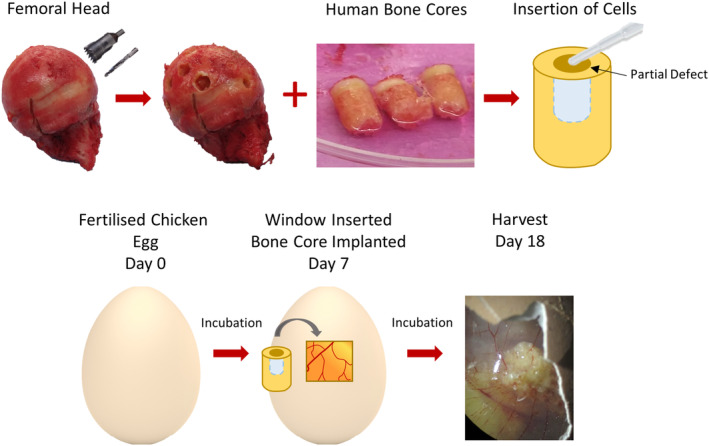
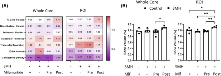
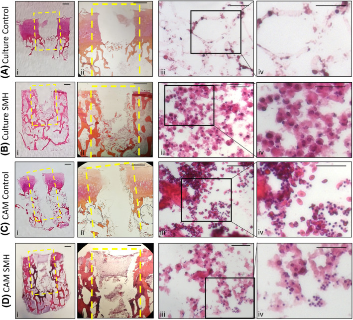
Osteosarcoma is the most common primary bone cancer, occurring frequently in children and young adults. Patients are treated with surgery and multi-agent chemotherapy, and despite the introduction of mifamurtide in 2011, there has been little improvement in survival for decades. 3-dimensional models offer the potential to understand the complexity of the osteosarcoma tumor microenvironment and aid in developing new treatment approaches. An osteosarcoma 3D bone core model was developed using human trabecular bone and the chorioallantoic membrane (CAM), to form a functioning vasculature. A tri-culture of cells, stromal cells, macrophages, and the Saos-2 osteosarcoma cell line, were implanted into this model to simulate components of the tumor microenvironment, and mifamurtide was tested in this context. Immunohistochemistry and micro-CT were performed to assess phenotypic and structural effects of implantation. Successful integration and angiogenesis of the bone cores were observed after incubation on the CAM. The 3D bone model also showed similar characteristics to osteosarcoma patient samples including CD68 and CD105 expression. Incubating bone cores with mifamurtide induced a reduction of cellular markers and an increase in bone volume. This 3D bone core model has the potential to investigate osteosarcoma tumor microenvironment and provides a representative model for evaluation of novel therapies.
骨肉瘤是最常见的原发性骨癌,在儿童和年轻人中频繁发生。患者接受手术和多药联合化疗,尽管2011年引入了米伐木肽,但几十年来生存率几乎没有改善。三维模型为理解骨肉瘤肿瘤微环境的复杂性以及开发新的治疗方法提供了可能。利用人松质骨和绒毛尿囊膜(CAM)构建了一个骨肉瘤三维骨芯模型,以形成功能性脉管系统。将细胞、基质细胞、巨噬细胞和Saos-2骨肉瘤细胞系进行共培养,植入该模型以模拟肿瘤微环境的组成部分,并在此背景下对米伐木肽进行了测试。进行免疫组织化学和显微CT以评估植入的表型和结构效应。在CAM上孵育后观察到骨芯的成功整合和血管生成。该三维骨模型还显示出与骨肉瘤患者样本相似的特征,包括CD68和CD105表达。用米伐木肽孵育骨芯可导致细胞标志物减少和骨体积增加。这种三维骨芯模型有潜力研究骨肉瘤肿瘤微环境,并为评估新疗法提供一个代表性模型。