Okazaki Takashi, Niwa Tetsu, Yoshida Ryoichi, Sorimachi Takatoshi, Hashimoto Jun
Department of Diagnostic Radiology, Tokai University School of Medicine, 143 Shimokasuya, Isehara 259-1193, Japan.
Department of Radiology, Tokai University Hospital, 143 Shimokasuya, Isehara 259-1193, Japan.
Tomography. 2024 Nov 21;10(12):1867-1880. doi: 10.3390/tomography10120136.
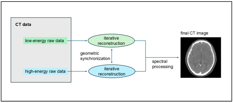
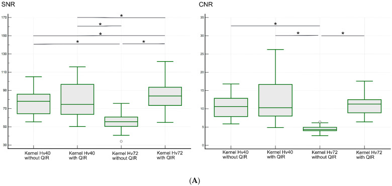
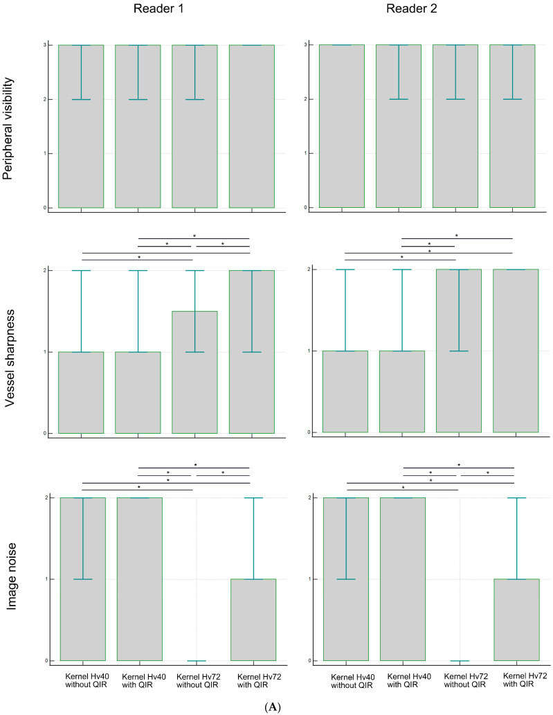
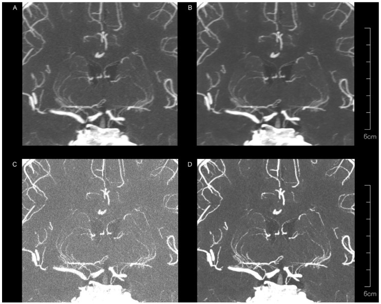
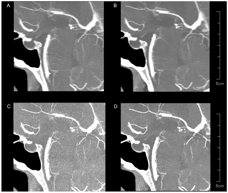
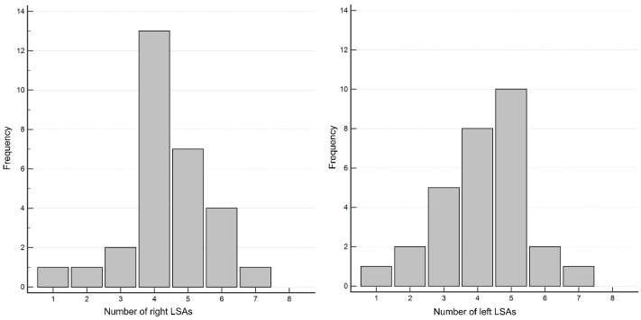
Photon-counting detector computed tomography (PCD-CT) offers energy-resolved CT data with enhanced resolution, reduced electronic noise, and improved tissue contrast. This study aimed to evaluate the visibility of intracranial perforating arteries on ultra-high-resolution (UHR) CT angiography (CTA) on PCD-CT. A retrospective analysis of intracranial UHR PCD-CTA was performed for 30 patients. The image quality from four UHR PCD-CTA reconstruction methods [kernel Hv40 and Hv72, with and without quantum iterative reconstruction (QIR)] was assessed for the lenticulostriate arteries (LSAs) and pontine arteries (PAs). A subjective evaluation included peripheral visibility, vessel sharpness, and image noise, while objective analysis focused on the signal-to-noise ratio (SNR) and contrast-to-noise ratio (CNR). Peripheral LSAs were well visualized across all reconstruction methods, with no significant differences between them. Vessel sharpness and image noise varied significantly ( < 0.0001); sharper LSAs and more noise were seen with kernel Hv72 compared to kernel Hv40 ( < 0.05). A similar pattern was observed for PAs, though peripheral visibility was lower than that for LSAs. The SNR and CNR were the highest in the presence of kernel Hv72 with QIR, and lowest with kernel Hv72 without QIR, compared to kernel Hv40 ( < 0.05). UHR PCD-CTA provided a good visualization of the intracranial perforating arteries, particularly LSAs. The vessel sharpness and image noise varied by reconstruction method, in which kernel Hv72 with QIR offered the optimal visualization.
光子计数探测器计算机断层扫描(PCD-CT)可提供能量分辨的CT数据,具有更高的分辨率、更低的电子噪声和更好的组织对比度。本研究旨在评估颅内穿支动脉在PCD-CT上的超高分辨率(UHR)CT血管造影(CTA)中的显示情况。对30例患者进行了颅内UHR PCD-CTA的回顾性分析。对四种UHR PCD-CTA重建方法[内核Hv40和Hv72,有无量子迭代重建(QIR)]的图像质量进行了评估,观察豆纹动脉(LSA)和脑桥动脉(PA)。主观评估包括外周可见性、血管清晰度和图像噪声,而客观分析则侧重于信噪比(SNR)和对比噪声比(CNR)。所有重建方法对外周LSA均显示良好,它们之间无显著差异。血管清晰度和图像噪声差异显著(<0.0001);与内核Hv40相比,内核Hv72显示的LSA更清晰,但噪声更多(<0.05)。PA也观察到类似模式,尽管外周可见性低于LSA。与内核Hv40相比,内核Hv72联合QIR时SNR和CNR最高,内核Hv72不联合QIR时最低(<0.05)。UHR PCD-CTA能很好地显示颅内穿支动脉,尤其是LSA。血管清晰度和图像噪声因重建方法而异,其中内核Hv72联合QIR时显示最佳。