Kamel Asmaa Khalf, Farag Naglaa M, Allam Emad, Khaled Mohamed, Ismail Doaa Elzaeem
Department of Clinical Pathology, Minia University Faculty of Medicine, Minia, EGY.
Department of Cardiology, Minia University Faculty of Medicine, Minia, EGY.
Cureus. 2024 Nov 28;16(11):e74670. doi: 10.7759/cureus.74670. eCollection 2024 Nov.
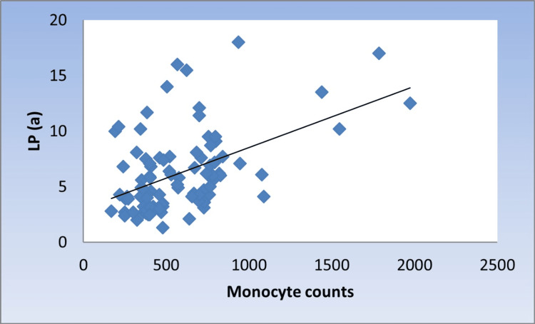
Introduction Many studies have supported inflammation as a mediator of lipoprotein (a) (Lp(a)) induced increase in cardiovascular disease risk, as it has pro-inflammatory effects on endothelial cells and monocytes. Aim This study aims to correlate Lp(a) level with different monocyte subsets in coronary atherosclerotic patients with different severity. Method The study included 60 patients with a mean age of 53.1 ± 10.5 diagnosed as coronary atherosclerotic patients by coronary angiography. Lp levels were measured using enzyme-linked immunosorbent assay (ELISA), while blood counts and monocyte subsets were analyzed by flow cytometry, and 30 apparently healthy individuals were included as the control group. Results Patients showed significantly higher median monocytic %, Lp(a), and higher C-reactive protein (CRP) values than the control group. Patients were subdivided into two groups: normal Lp(a) < 6.2 mg/dL (n = 24) and hyperlipoproteinemia(a) (hyper Lp(a)) ≥ 6.2 mg/dL (n = 36). Patients with hyper Lp(a) had higher non-classical monocytes (31.5% vs. 20%). Coronary atherosclerosis severity was associated with higher Lp(a) levels as well as non-classical monocytes; patients with mild atherosclerosis showed the highest classical and intermediate subset levels. While for a non-classical subset, patients with severe atherosclerosis showed the highest median level. A significant moderate positive correlation between Lp(a) and monocyte counts, as well as monocyte-lymphocyte (M/L) index and non-classical monocytes, was found. Conclusions Hyper Lp(a) and increased count of non-classical monocytes are significantly increased with disease progression (triple-vessel coronary disease risk). These results suggest that the expansion of non-classical monocytes is a cardiovascular disease (CVD) risk and predictor for disease severity. Strategies targeting inflammatory monocytes may slow CVD progression.
引言 许多研究支持炎症作为脂蛋白(a) [Lp(a)] 诱导心血管疾病风险增加的介质,因为它对内皮细胞和单核细胞具有促炎作用。目的 本研究旨在探讨不同严重程度的冠状动脉粥样硬化患者中Lp(a)水平与不同单核细胞亚群之间的相关性。方法 本研究纳入了60例平均年龄为53.1±10.5岁的患者,这些患者经冠状动脉造影诊断为冠状动脉粥样硬化患者。采用酶联免疫吸附测定(ELISA)法测定Lp水平,同时通过流式细胞术分析血细胞计数和单核细胞亚群,并纳入30名明显健康的个体作为对照组。结果 患者的单核细胞百分比中位数、Lp(a)以及C反应蛋白(CRP)值均显著高于对照组。患者被分为两组:正常Lp(a)<6.2mg/dL(n=24)和高脂蛋白血症(a) [高Lp(a)]≥6.2mg/dL(n=36)。高Lp(a)患者的非经典单核细胞比例更高(31.5%对20%)。冠状动脉粥样硬化的严重程度与较高的Lp(a)水平以及非经典单核细胞相关;轻度动脉粥样硬化患者的经典和中间亚群水平最高。而对于非经典亚群,重度动脉粥样硬化患者的中位数水平最高。发现Lp(a)与单核细胞计数、单核细胞-淋巴细胞(M/L)指数和非经典单核细胞之间存在显著的中度正相关。结论 随着疾病进展(三支血管冠状动脉疾病风险),高Lp(a)和非经典单核细胞计数显著增加。这些结果表明,非经典单核细胞的扩增是心血管疾病(CVD)的风险因素和疾病严重程度的预测指标。针对炎性单核细胞的策略可能会减缓CVD的进展。