Lapras Benjamine, Merienne Camille, Eynaud Emma, Usseglio Léa, Marchand Chloé, Médina Mathieu, Kolenda Camille, Briot Thomas, Laurent Frédéric, Pirot Fabrice
Pharmacy Department, Hospices Civils de Lyon, Hôpital E. Herriot, Plateforme FRIPHARM, 69437, Lyon, France.
Tissue Biology and Therapeutic Engineering Laboratory (LBTI), CNRS UMR 5305, 69007, Lyon, France.
Sci Rep. 2024 Dec 30;14(1):31629. doi: 10.1038/s41598-024-79478-w.
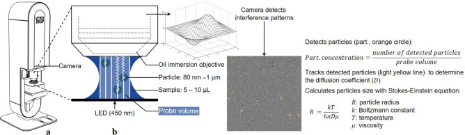
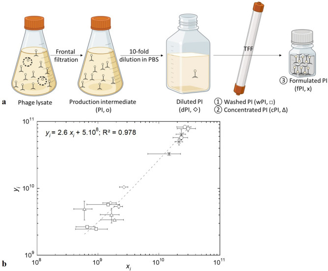
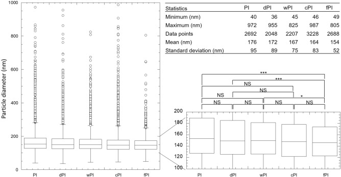
Phage therapy uses viruses (phages) against antibiotic resistance. Tailoring treatments to specific patient strains requires stocks of various highly concentrated purified phages. It, therefore, faces challenges: titration duration and specificity to a phage/bacteria couple; purification affecting stability; and highly concentrated suspensions tending to aggregate. To address these challenges, interferometric light microscopy (ILM), characterising particles (size, concentration, and visual homogeneity) within minutes, was applied herein to anti-Staphylococcus aureus myovirus phage suspensions. Particle concentration was linearly correlated with phage infectious titre (R > 0.97, slope: 3 particles/plaque forming units (PFU)) at various degrees of purification, allowing to approximate the infectious titre for suspensions ≥ 3 × 10 PFU/mL, thereby encompassing most therapeutic doses. Purification narrowed and homogenised particle distribution while maintaining therapeutic concentrations. When compared to dynamic light scattering, electrophoretic mobility, and UV/Visible-spectroscopy, ILM best detected aggregates according to our homemade scoring. Although ILM has certain limitations, such as the inability to detect podoviruses (hydrodynamic diameter < 80 nm), or to measure particles in low-concentrated suspensions (< 10 particles/mL), the present proof-of-concept positions this technique as a valuable quality control tool, as a complement to titration rather than a replacement for this technique, for phage suspensions, paving the way for further investigations.
噬菌体疗法利用病毒(噬菌体)来对抗抗生素耐药性。针对特定患者菌株定制治疗方案需要各种高浓度纯化噬菌体的储备。因此,它面临着挑战:滴定持续时间以及噬菌体/细菌对的特异性;纯化影响稳定性;以及高浓度悬浮液容易聚集。为应对这些挑战,本文将干涉光显微镜(ILM)应用于抗金黄色葡萄球菌肌病毒噬菌体悬浮液,该技术可在数分钟内对颗粒(大小、浓度和视觉均匀性)进行表征。在不同纯化程度下,颗粒浓度与噬菌体感染滴度呈线性相关(R > 0.97,斜率:3个颗粒/噬菌斑形成单位(PFU)),这使得对于≥3×10 PFU/mL的悬浮液能够近似估算感染滴度,从而涵盖了大多数治疗剂量。纯化使颗粒分布变窄并均匀化,同时保持治疗浓度。与动态光散射、电泳迁移率和紫外/可见光谱相比,根据我们自制的评分标准,ILM能最好地检测到聚集体。尽管ILM有一定局限性,如无法检测短尾噬菌体(流体动力学直径<80 nm),或无法测量低浓度悬浮液(<10个颗粒/mL)中的颗粒,但本概念验证将该技术定位为一种有价值的质量控制工具,作为噬菌体悬浮液滴定的补充而非替代该技术,为进一步研究铺平了道路。