Liu Xi, Zhang Xi, Wang Dongxu, Cao Ya, Zhang Ling, Li Zhonghua, Zhang Qin, Shen Yu, Lu Xian, Fan Keyu, Liu Mingxia, Wei Jingqiu, Hu Siping, Liu He
Department of Anesthesiology & Clinical Research Center for Anesthesia and Perioperative Medicine & Key Laboratory of Anesthesia and Analgesia Application Technology, Huzhou Central Hospital, The Fifth School of Clinical Medicine of Zhejiang Chinese Medical University, Huzhou, China.
Department of Anesthesiology & Clinical Research Center for Anesthesia and Perioperative Medicine & Key Laboratory of Anesthesia and Analgesia Application Technology, Huzhou Central Hospital, The Affiliated Central Hospital of Huzhou University, Huzhou, China.
Brain Behav. 2025 Jan;15(1):e70218. doi: 10.1002/brb3.70218.
Pain is a prevalent comorbidity in numerous clinical conditions and causes suffering; however, the mechanism of pain is intricate, and the neural circuitry underlying pain in the brain remains incompletely elucidated. More research into the perception and modulation of pain within the central nervous system is essential. The nucleus accumbens (NAc) plays a pivotal role in the regulation of animal behavior, and extensive research has unequivocally demonstrated its significant involvement in the occurrence and development of pain. NAc receives projections from various other neural nuclei within the brain, including the paraventricular nucleus of the thalamus (PVT). In this experiment, we demonstrate that the specific glutamatergic neural circuit projection from PVT to NAc (PVT→NAc) is implicated in the modulation of inflammatory pain in mice.
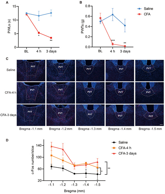
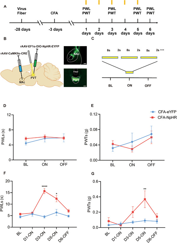
We compared the difference in pain thresholds between complete Freund's adjuvant (CFA)-induced inflammatory pain models and controls. Then in a well-established mouse model of CFA-induced inflammatory pain, immunofluorescence staining was utilized to evaluate changes in c-Fos protein expression within PVT neurons. To investigate the role of PVT→NAc in the modulation of pain, we used optogenetics to modulate this neural circuit, and nociceptive behavioral tests were employed to investigate the functional role of the PVT→NAc circuit in the modulation of inflammatory pain.
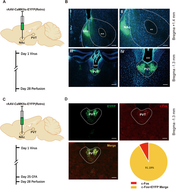
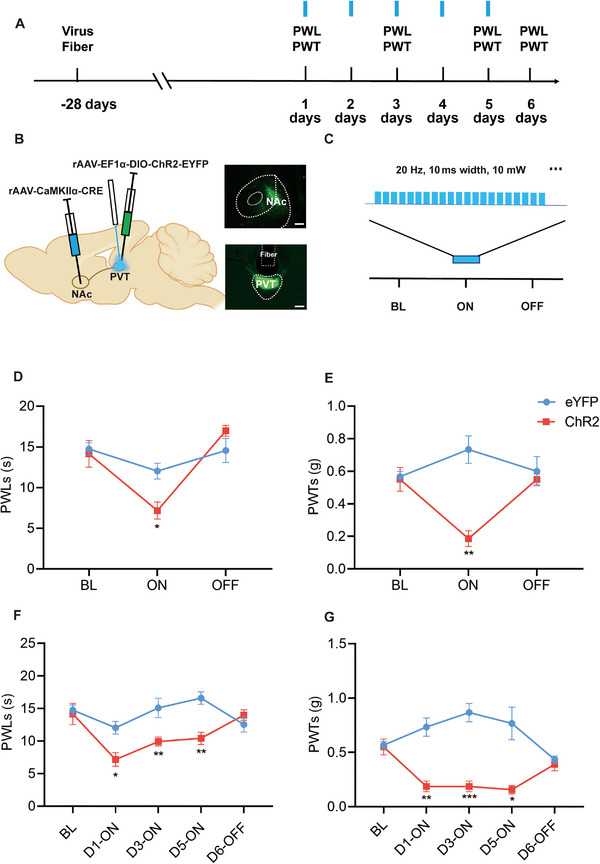
In the mice with the inflammatory pain group, both the paw withdrawal latencies (PWLs) and paw withdrawal thresholds (PWTs) of the right hind paw were decreased compared to the control group. In addition, compared to the control group, CFA-induced inflammatory pain led to increased c-Fos protein expression in PVT, which means that some of the neurons in this area of the brain region have been activated. Following the injection of retrograde transport fluorescent-labeled virus into NAc, glutamatergic neurons projecting from the PVT to NAc were observed, confirming the projection relationship between PVT and NAc. In the experiments in optogenetic regulation, normal mice exhibited pain behavior when the PVT→NAc circuit was stimulated by a 473 nm blue laser, resulting in decreased PWLs and PWTs compared to the control group, which means activating this neural circuit can lead to painful behaviors. In the CFA-induced pain group, inhibition of the PVT→NAc circuit by a 589 nm yellow laser alleviated pain behavior, leading to increased PWLs and PWTs compared to the control group, representing the fact that inhibition of this neural circuit relieves pain behaviors.
The findings unveil a pivotal role of the PVT→NAc circuit in modulating inflammatory pain induced by CFA in mice.
疼痛是众多临床病症中普遍存在的合并症,会带来痛苦;然而,疼痛机制错综复杂,大脑中疼痛的神经回路仍未完全阐明。对中枢神经系统内疼痛的感知与调节进行更多研究至关重要。伏隔核(NAc)在动物行为调节中起关键作用,大量研究已明确表明其在疼痛的发生和发展中具有重要作用。NAc接收来自大脑内其他各种神经核的投射,包括丘脑室旁核(PVT)。在本实验中,我们证明从PVT到NAc的特定谷氨酸能神经回路投射(PVT→NAc)与小鼠炎症性疼痛的调节有关。
我们比较了完全弗氏佐剂(CFA)诱导的炎症性疼痛模型与对照组之间疼痛阈值的差异。然后,在成熟的CFA诱导的炎症性疼痛小鼠模型中,利用免疫荧光染色评估PVT神经元内c-Fos蛋白表达的变化。为了研究PVT→NAc在疼痛调节中的作用,我们使用光遗传学来调节该神经回路,并采用伤害性行为测试来研究PVT→NAc回路在炎症性疼痛调节中的功能作用。
在炎症性疼痛组小鼠中,与对照组相比,右后爪的爪退缩潜伏期(PWLs)和爪退缩阈值(PWTs)均降低。此外,与对照组相比,CFA诱导的炎症性疼痛导致PVT中c-Fos蛋白表达增加,这意味着该脑区的一些神经元已被激活。向NAc注射逆行运输荧光标记病毒后,观察到从PVT投射到NAc的谷氨酸能神经元,证实了PVT与NAc之间的投射关系。在光遗传学调节实验中,当用473nm蓝色激光刺激PVT→NAc回路时,正常小鼠表现出疼痛行为,与对照组相比,PWLs和PWTs降低,这意味着激活该神经回路会导致疼痛行为。在CFA诱导的疼痛组中,用589nm黄色激光抑制PVT→NAc回路可减轻疼痛行为,与对照组相比,PWLs和PWTs增加,这表明抑制该神经回路可缓解疼痛行为。
这些发现揭示了PVT→NAc回路在调节小鼠CFA诱导的炎症性疼痛中的关键作用。