Guo Wei, Zhou Bolun, Dou Lizhou, Guo Lei, Li Yong, Qin Jianjun, Wang Zhen, Huai Qilin, Xue Xuemin, Li Yin, Ying Jianming, Xue Qi, Gao Shugeng, He Jie
Department of Thoracic Surgery, National Cancer Center/National Clinical Research Center for Cancer/Cancer Hospital, Chinese Academy of Medical Sciences and Peking Union Medical College, Beijing, China.
Key Laboratory of Minimally Invasive Therapy Research for Lung Cancer, Chinese Academy of Medical Sciences, Beijing, China.
Exp Mol Med. 2025 Feb;57(1):59-71. doi: 10.1038/s12276-024-01369-x. Epub 2025 Jan 1.
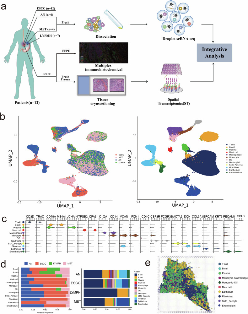
Esophageal squamous cell carcinoma (ESCC) patients often face a grim prognosis due to lymph node metastasis. However, a comprehensive understanding of the cellular and molecular characteristics of metastatic lymph nodes in ESCC remains elusive. In this study involving 12 metastatic ESCC patients, we employed single-cell sequencing, spatial transcriptomics (ST), and multiplex immunohistochemistry (mIHC) to explore the spatial and molecular attributes of primary tumor samples, adjacent tissues, metastatic and non-metastatic lymph nodes. The analysis of 161,333 cells revealed specific subclusters of epithelial cells that were significantly enriched in metastatic lymph nodes, suggesting pro-metastatic characteristics. Furthermore, stromal cells in the tumor microenvironment, including MMP3IL24 fibroblasts, APLN endothelial cells, and CXCL12 pericytes, were implicated in ESCC metastasis through angiogenesis, collagen production, and inflammatory responses. Exhausted CD8 T cells in a cycling status were notably prevalent in metastatic lymph nodes, indicating their potential role in facilitating metastasis. We identified distinct cell-cell communication networks and specific ligand-receptor pathways. Our findings were validated through a spatial transcriptome map and mIHC. This study enhances our comprehension of the cellular and molecular aspects of metastatic lymph nodes in ESCC patients, offering potential insights into novel therapeutic strategies for these individuals.
食管鳞状细胞癌(ESCC)患者常因淋巴结转移而面临严峻的预后。然而,对ESCC转移性淋巴结的细胞和分子特征的全面了解仍然难以捉摸。在这项涉及12例转移性ESCC患者的研究中,我们采用单细胞测序、空间转录组学(ST)和多重免疫组化(mIHC)来探索原发性肿瘤样本、相邻组织、转移性和非转移性淋巴结的空间和分子属性。对161,333个细胞的分析揭示了上皮细胞的特定亚群,这些亚群在转移性淋巴结中显著富集,表明具有促转移特征。此外,肿瘤微环境中的基质细胞,包括MMP3IL24成纤维细胞、APLN内皮细胞和CXCL12周细胞,通过血管生成、胶原蛋白产生和炎症反应参与ESCC转移。处于循环状态的耗竭CD8 T细胞在转移性淋巴结中显著普遍,表明它们在促进转移中的潜在作用。我们确定了不同的细胞间通讯网络和特定的配体-受体途径。我们的发现通过空间转录组图谱和mIHC得到验证。这项研究增强了我们对ESCC患者转移性淋巴结的细胞和分子方面的理解,为这些患者的新治疗策略提供了潜在的见解。