Liu Xianwang, Han Tao, Wang Yuzhu, Liu Hong, Deng Juan, Xue Caiqiang, Li Shenglin, Zhou Junlin
Department of Radiology, Lanzhou University Second Hospital, Cuiyingmen No.82, Chengguan District, Lanzhou, 730030, People's Republic of China.
Second Clinical School, Lanzhou University, Lanzhou, People's Republic of China.
Cancer Imaging. 2024 Dec 31;24(1):173. doi: 10.1186/s40644-024-00820-6.
To assess and compare the diagnostic efficiency of histogram analysis of monochromatic and iodine images derived from spectral CT in predicting Ki-67 expression in gastric gastrointestinal stromal tumors (gGIST).
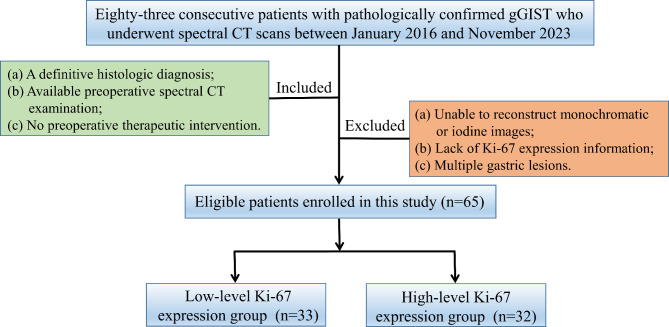
Sixty-five patients with gGIST who underwent spectral CT were divided into a low-level Ki-67 expression group (LEG, Ki-67 < 10%, n = 33) and a high-level Ki-67 expression group (HEG, Ki-67 ≥ 10%, n = 32). Conventional CT features were extracted and compared. Histogram parameters were extracted from monochromatic and iodine images, respectively. The diagnostic efficiency of the histogram parameters from monochromatic and iodine images was assessed and compared between the two groups. Spearman's correlation analysis was used to correlate histogram parameters with Ki-67 expression.
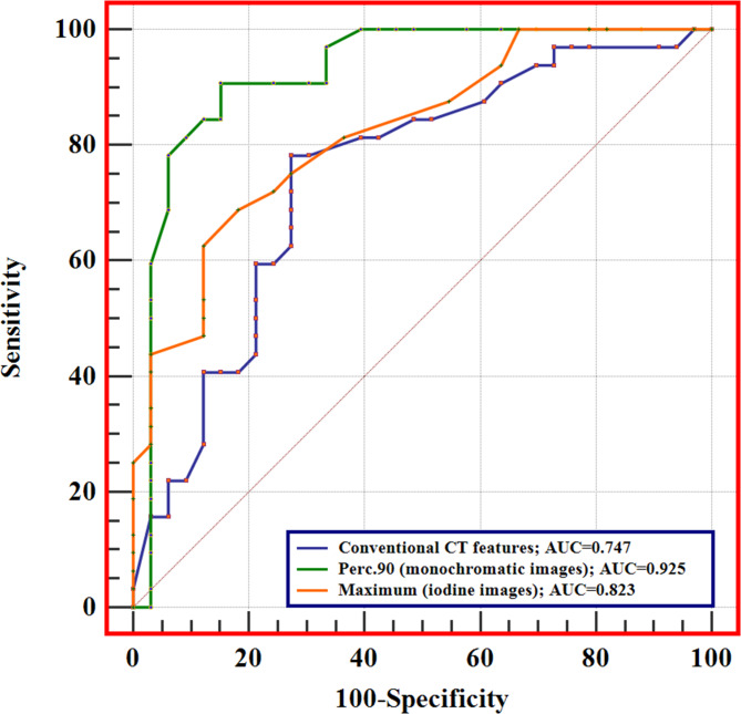
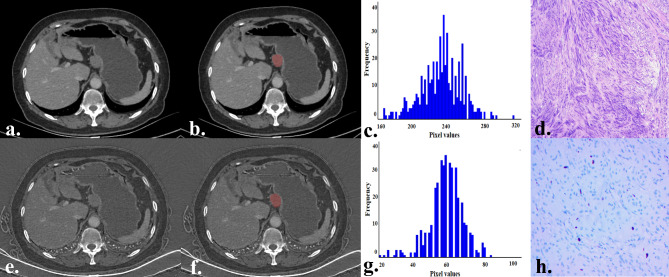
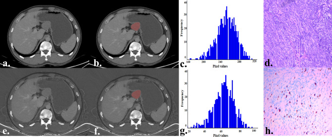
The HEG was more likely to present with an irregular shape and a larger size than the LEG (all p < 0.05). Regarding histogram parameters, the HEG showed higher maximum, mean, Perc.10, Perc.25, Perc.50, Perc.75, Perc.90, Perc.99, SD, variance, and CV of monochromatic images; higher maximum, Perc.99, and entropy of iodine images, compared with the LEG (all p < 0.003125). ROC analysis showed that significant histogram parameters of monochromatic and iodine images allowed for effective differentiation between LEG and HEG. DeLong's test showed that the diagnostic efficiency of histogram parameters in monochromatic images (Perc.90) was superior to that of iodine images (maximum) (p = 0.010). A positive correlation was observed between the significant histogram parameters and Ki-67 expression (all p < 0.05).
Both histogram analysis of monochromatic and iodine images derived from spectral CT can predict Ki-67 expression in gGIST, and the diagnostic efficacy of monochromatic images is superior to iodine images.
评估并比较光谱CT单色图像和碘图像的直方图分析在预测胃胃肠道间质瘤(gGIST)中Ki-67表达方面的诊断效率。
65例接受光谱CT检查的gGIST患者被分为低水平Ki-67表达组(LEG,Ki-67<10%,n = 33)和高水平Ki-67表达组(HEG,Ki-67≥10%,n = 32)。提取并比较常规CT特征。分别从单色图像和碘图像中提取直方图参数。评估并比较两组中单色图像和碘图像直方图参数的诊断效率。采用Spearman相关分析将直方图参数与Ki-67表达进行关联。
与LEG组相比,HEG组更易出现不规则形状且尺寸更大(所有p<0.05)。关于直方图参数,与LEG组相比,HEG组在单色图像中的最大值、平均值、第10百分位数、第25百分位数、第50百分位数、第75百分位数、第90百分位数、第99百分位数、标准差、方差和变异系数更高;在碘图像中的最大值、第99百分位数和熵更高(所有p<0.003125)。ROC分析表明,单色图像和碘图像的显著直方图参数能够有效区分LEG组和HEG组。DeLong检验表明,单色图像直方图参数(第90百分位数)的诊断效率优于碘图像(最大值)(p = 0.010)。显著直方图参数与Ki-67表达之间存在正相关(所有p<0.05)。
光谱CT的单色图像和碘图像的直方图分析均可预测gGIST中的Ki-67表达,且单色图像的诊断效能优于碘图像。