Droby Amgad, Yoffe-Vasiliev Avital, Atias Daniel, Fraser Kyle B, Mabrouk Omar S, Omer Nurit, Bar-Shira Anat, Gana-Weisz Mali, Goldstein Orly, Artzi Moran, Ben Bashat Dafna, Alcalay Roy N, Orr-Urtreger Avi, Shirvan Julia C, Cedarbaum Jesse M, Giladi Nir, Mirelman Anat, Thaler Avner
Movement Disorders Unit, Neurological Institute, Tel Aviv Medical Center, Tel Aviv, Israel.
Laboratory for Early Markers of Neurodegeneration, Neurological Institute, Tel Aviv Medical Center, Tel Aviv, Israel.
NPJ Parkinsons Dis. 2025 Jan 3;11(1):7. doi: 10.1038/s41531-024-00854-4.
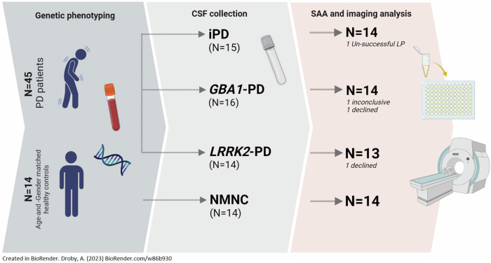
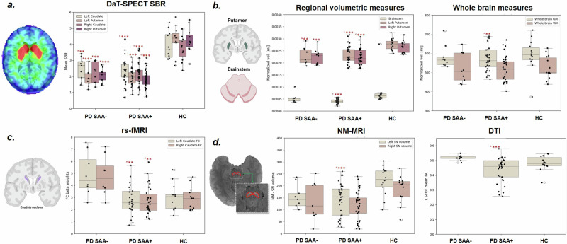
Alpha-synuclein (αS) aggregation is a widely regarded hallmark of Parkinson's disease (PD) and can be detected through synuclein amplification assays (SAA). This study investigated the association between cerebrospinal fluid (CSF) radiological measures in 41 PD patients (14 iPD, 14 GBA1-PD, 13 LRRK2-PD) and 14 age-and-sex-matched healthy controls. Quantitative measures including striatal binding ratios (SBR), whole-brain and deep gray matter volumes, neuromelanin-MRI (NM-MRI), functional connectivity (FC), and white matter (WM) diffusion-tensor imaging (DTI) were calculated. Nine LRRK2-PD patients were SAA-negative (PD-SAA-). PD-SAA+ patients showed lower whole-brain gray matter, putamenal, brainstem, and substantia nigra volumes, reduced FC in the left caudate, and lower fractional anisotropy in the left fronto-occipital fasciculus compared to PD-SAA-. Taken together, αS aggregation was observed in iPD, GBA1-PD, and 38% of LRRK2-PD patients, and this was associated with reduced regional brain volumes, altered caudal FC, and SBRs. These changes were less pronounced in PD-SAA-, possibly suggesting a milder neurodegenerative process.
α-突触核蛋白(αS)聚集是帕金森病(PD)广泛认可的一个标志,可通过突触核蛋白扩增检测(SAA)来检测。本研究调查了41例PD患者(14例散发性帕金森病患者(iPD)、14例GBA1基因相关帕金森病患者(GBA1-PD)、13例富亮氨酸重复激酶2基因相关帕金森病患者(LRRK2-PD))与14名年龄和性别匹配的健康对照者脑脊液(CSF)的影像学指标之间的关联。计算了包括纹状体结合率(SBR)、全脑和深部灰质体积、神经黑色素磁共振成像(NM-MRI)、功能连接(FC)以及白质(WM)扩散张量成像(DTI)等定量指标。9例LRRK2-PD患者SAA检测为阴性(PD-SAA-)。与PD-SAA-患者相比,PD-SAA+患者的全脑灰质、壳核、脑干和黑质体积更小,左侧尾状核的FC降低,左侧额枕束的分数各向异性更低。总体而言,在iPD、GBA1-PD以及38%的LRRK2-PD患者中观察到了αS聚集,这与脑区体积减小、尾侧FC改变以及SBR有关。这些变化在PD-SAA-患者中不太明显,这可能表明神经退行性变过程较为轻微。