Zucker Ben, Dharan Raviv, Wang Dongju, Yu Li, Sorkin Raya, Kozlov Michael M
Department of Physiology and Pharmacology, Faculty of Medical and Health Sciences, Tel Aviv University, Tel Aviv, Israel; Center for Physics and Chemistry of Living Systems, Tel Aviv University, Tel Aviv, Israel.
School of Chemistry, Faculty of Exact Sciences, Tel Aviv University, Tel Aviv, Israel; Center for Physics and Chemistry of Living Systems, Tel Aviv University, Tel Aviv, Israel.
Biophys J. 2025 Feb 18;124(4):604-619. doi: 10.1016/j.bpj.2024.12.029. Epub 2025 Jan 3.
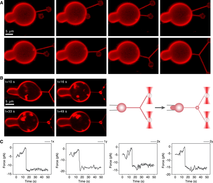
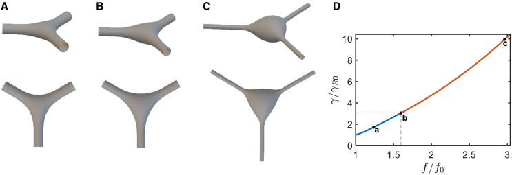
Migrasomes, the vesicle-like membrane microstructures, arise on the retraction fibers (RFs), the branched nanotubules pulled out of cell plasma membranes during cell migration and shaped by membrane tension. Migrasomes form in two steps: a local RF bulging is followed by a protein-dependent stabilization of the emerging spherical bulge. Here, we addressed theoretically and experimentally the previously unexplored mechanism of bulging of membrane tubular systems. We assumed that the bulging could be driven by increases in membrane tension and experimentally verified this hypothesis in live-cell and biomimetic systems. We exposed RF-generating live cells to a hypotonic medium, which produced water flows into the cells and a related increase in the membrane tension. We observed the formation of migrasome-like bulges with a preferential location in the RF branching sites. Next, we developed a biomimetic system of three membrane tubules pulled out of a giant plasma membrane vesicle (GPMV), connected by a junction, and subjected to pulling forces controlled by the GPMV membrane tension. An abrupt increase in the GPMV tension resulted in the generation of migrasome-like bulges mainly in the junctions. To understand the physical forces behind these observations, we considered theoretically the mechanical energy of a membrane system consisting of a three-way tubular junction with emerging tubular arms subjected to membrane tension. Substantiating our experimental observations, the energy minimization predicted a tension increase to drive the formation of membrane bulges, preferably in the junction site, independently of the way of the tension application. We generalized the model to derive universal criteria of bulging in branched membrane tubules.
迁移小体是一种囊泡状的膜微结构,出现在收缩纤维(RFs)上,收缩纤维是细胞迁移过程中从细胞质膜拉出的分支纳米管,并由膜张力塑造而成。迁移小体的形成分为两个步骤:首先是局部RF凸起,随后是新生球形凸起的蛋白质依赖性稳定化。在这里,我们从理论和实验上探讨了膜管状系统凸起这一此前未被探索的机制。我们假设凸起可能是由膜张力增加驱动的,并在活细胞和仿生系统中通过实验验证了这一假设。我们将产生RF的活细胞暴露于低渗介质中,这会使水流进细胞并导致膜张力相应增加。我们观察到在RF分支位点优先形成了类似迁移小体的凸起。接下来,我们开发了一种仿生系统,该系统由从巨型质膜囊泡(GPMV)中拉出的三根膜管组成,通过一个连接点相连,并受到由GPMV膜张力控制的拉力作用。GPMV张力的突然增加导致主要在连接点处产生类似迁移小体的凸起。为了理解这些观察结果背后的物理力,我们从理论上考虑了一个膜系统的机械能,该膜系统由一个三叉管状连接点和受到膜张力作用的新生管状臂组成。能量最小化证实了我们的实验观察结果,即预测张力增加会驱动膜凸起的形成,最好是在连接点处,而与施加张力的方式无关。我们对模型进行了推广,以推导分支膜管中凸起的通用标准。