Ma Gaoyuan, Chan Jonathan M, Worthy Katrina H, Rosa Marcello G P, Atapour Nafiseh
Neuroscience Program, Biomedicine Discovery Institute and Department of Physiology, Monash University, Clayton, VIC, 3800, Australia.
Curr Res Neurobiol. 2024 Nov 28;8:100141. doi: 10.1016/j.crneur.2024.100141. eCollection 2025 Jun.
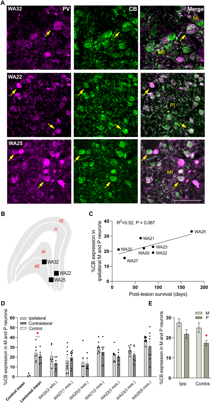
Lesions of the primary visual cortex (V1) cause retrograde neuronal degeneration, volume loss and neurochemical changes in the lateral geniculate nucleus (LGN). Here we characterised the timeline of these processes in adult marmoset monkeys, after various recovery times following unilateral V1 lesions. Observations in NeuN-stained sections obtained from animals with short recovery times (2, 3 or 14 days) showed that the volume and neuronal density in the LGN ipsilateral to the lesions were similar to those in the contralateral hemispheres. However, neuronal density in the lesion projection zone of LGN dropped rapidly thereafter, with approximately 50% of the population lost within a month post-lesion. This level of neuronal loss remained stable for over three years post-lesion. In comparison, shrinkage of the LGN volume progressed more gradually, not reaching a stable value until 6 months post lesion. We also determined the time course of the expression of the calcium-binding protein calbindin (CB) in magnocellular (M) and parvocellular (P) layer neurons, a form of neurochemical plasticity previously reported to be triggered by V1 lesions. We found that CB expression could be detected in surviving M and P neurons as early as two weeks after lesion, with the percentage of neurons showing this neurochemical phenotype gradually increasing over 6 months. Thus, neurochemical change precedes neuronal degeneration, suggesting it may be linked to a protective mechanism. This study highlights the limited time window for any possible interventions aimed at reducing secondary neuronal loss in the visual afferent pathways following damage to V1.
初级视觉皮层(V1)损伤会导致外侧膝状体核(LGN)发生逆行性神经元变性、体积减小和神经化学变化。在此,我们对成年狨猴单侧V1损伤后不同恢复时间的这些过程的时间进程进行了表征。对恢复时间较短(2、3或14天)的动物的NeuN染色切片观察表明,损伤同侧LGN的体积和神经元密度与对侧半球相似。然而,此后LGN损伤投射区域的神经元密度迅速下降,损伤后一个月内约有50%的神经元丢失。这种神经元丢失水平在损伤后三年多保持稳定。相比之下,LGN体积的缩小进展较为缓慢,直到损伤后6个月才达到稳定值。我们还确定了大细胞(M)和小细胞(P)层神经元中钙结合蛋白钙视网膜蛋白(CB)表达的时间进程,CB是先前报道的一种由V1损伤触发的神经化学可塑性形式。我们发现,损伤后两周即可在存活的M和P神经元中检测到CB表达,显示这种神经化学表型的神经元百分比在6个月内逐渐增加。因此,神经化学变化先于神经元变性,表明其可能与一种保护机制有关。这项研究强调了针对V1损伤后视觉传入通路中继发性神经元丢失进行任何可能干预的有限时间窗口。