Xiang Chuanxi, Raghunathan VijayKrishna, Qiu Yubin, Mehta Manisha, Demirs John T, Grosskreutz Cynthia L, Wilson Christopher W, Prasanna Ganesh
Ophthalmology, Novartis Biomedical Research, Cambridge, MA, USA.
Acta Neuropathol Commun. 2025 Jan 6;13(1):3. doi: 10.1186/s40478-024-01880-2.
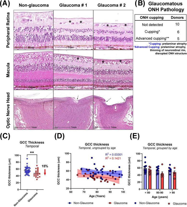
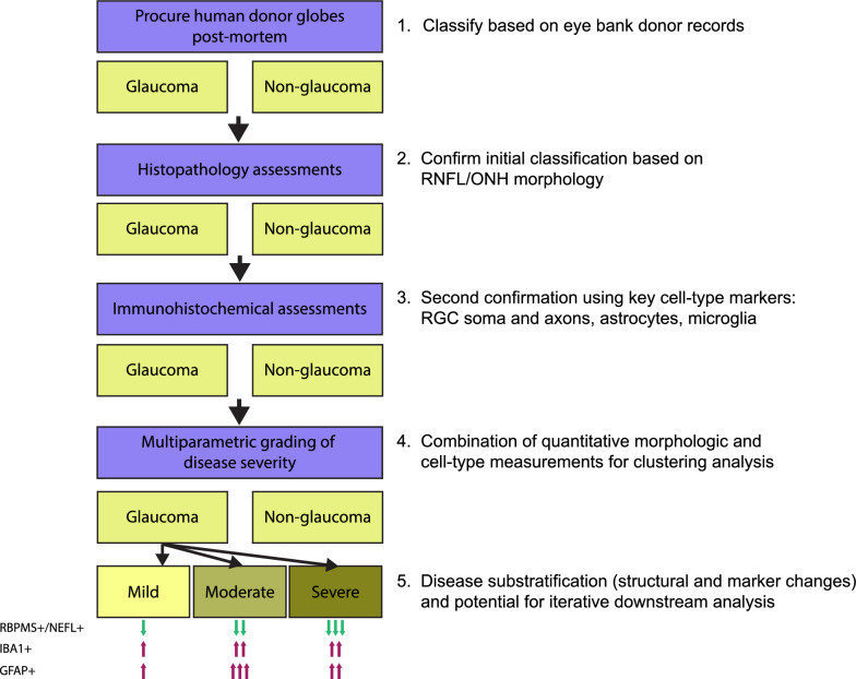
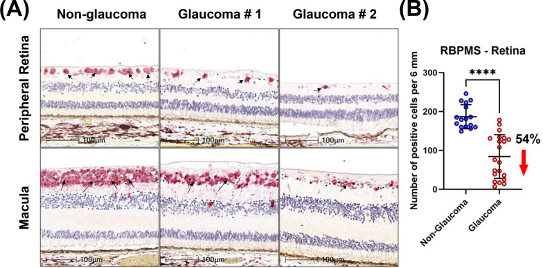
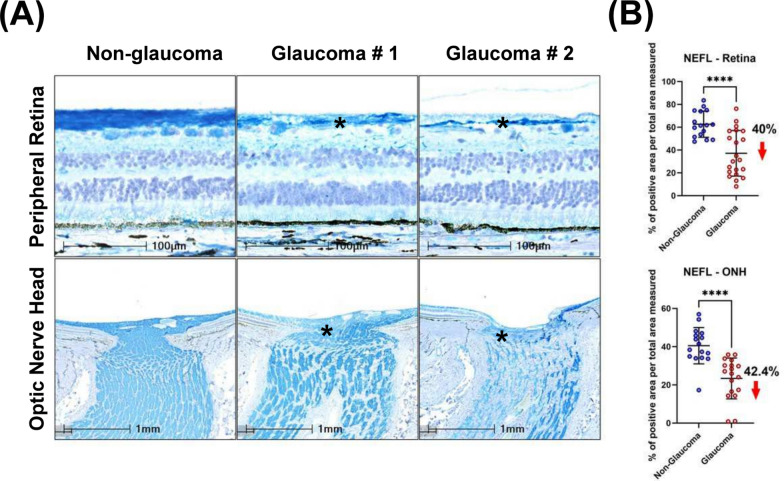
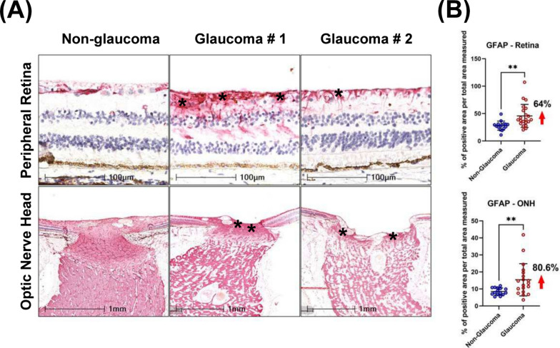
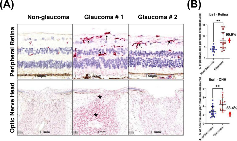
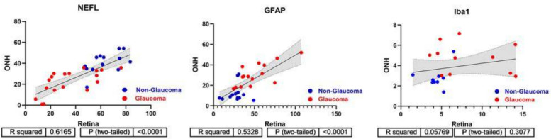
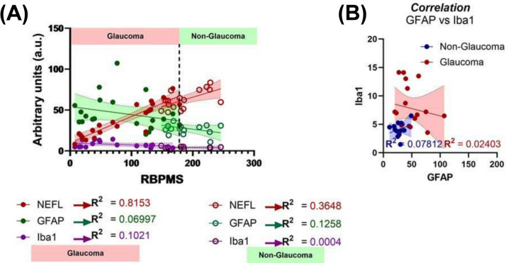
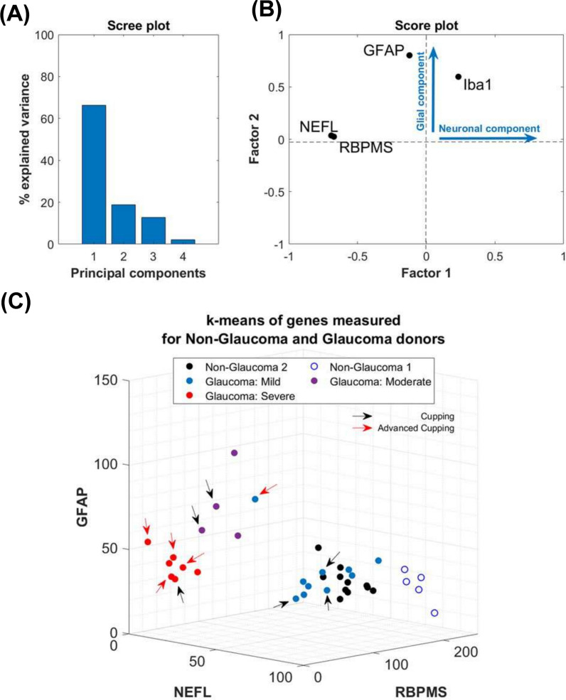
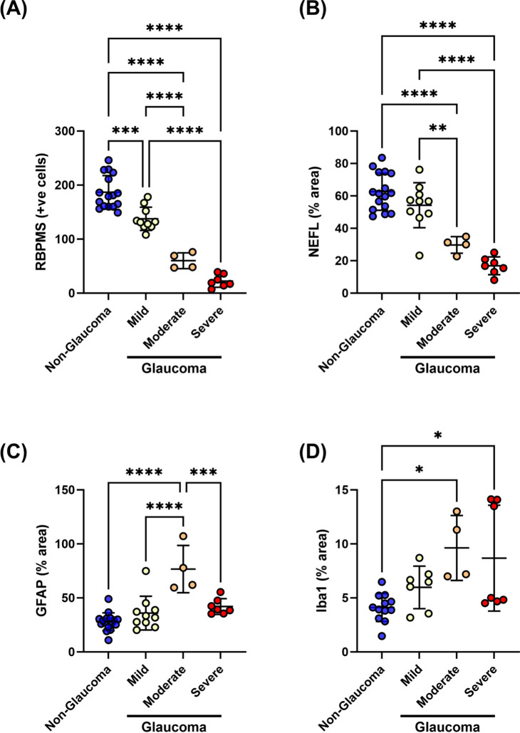
Neurodegeneration in glaucoma patients is clinically identified through longitudinal assessment of structure-function changes, including intraocular pressure, cup-to-disc ratios from fundus images, and optical coherence tomography imaging of the retinal nerve fiber layer. Use of human post-mortem ocular tissue for basic research is rising in the glaucoma field, yet there are challenges in assessing disease stage and severity, since tissue donations with informed consent are often unaccompanied by detailed pre-mortem clinical information. Further, the interpretation of disease severity based solely on anatomical and morphological assessments by histology can be affected by differences in death-to-preservation time and tissue processing. These are difficult confounders that cannot be easily controlled. As pathogenesis and molecular mechanisms can vary depending on the stage and severity of glaucoma, there is a need for the field to maximize use of donated tissue to better understand the molecular mechanisms of glaucoma and develop new therapeutic hypotheses. Further, there is a lack of consensus around the molecular RNA and protein markers that can be used to classify glaucoma severity. Here, we describe a multiparametric grading system that combines structural measurements of the retinal nerve fiber layer with linear regression and principal component analyses of molecular markers of retinal ganglion cells and glia (RBPMS, NEFL, IBA1 and GFAP) to stratify post-mortem glaucoma eyes by the severity of disease. Our findings show that a quantitative grading approach can stratify post-mortem glaucoma samples with minimal clinical histories into at least three severity groups and suggest that this type of approach may be useful for researchers aiming to maximize insights derived from eye bank donor tissue.
青光眼患者的神经退行性变是通过对结构-功能变化进行纵向评估来临床确定的,这些变化包括眼压、眼底图像的杯盘比以及视网膜神经纤维层的光学相干断层扫描成像。在青光眼领域,使用人类死后眼部组织进行基础研究的情况日益增多,但在评估疾病阶段和严重程度方面存在挑战,因为经知情同意的组织捐赠往往没有详细的生前临床信息。此外,仅基于组织学的解剖学和形态学评估来解释疾病严重程度可能会受到死亡至保存时间和组织处理差异的影响。这些是难以控制的混杂因素。由于青光眼的发病机制和分子机制可能因疾病阶段和严重程度而异,该领域需要最大限度地利用捐赠组织,以更好地了解青光眼的分子机制并提出新的治疗假设。此外,对于可用于对青光眼严重程度进行分类的分子RNA和蛋白质标志物,目前缺乏共识。在此我们描述了一种多参数分级系统,该系统将视网膜神经纤维层的结构测量与视网膜神经节细胞和神经胶质细胞分子标志物(RBPMS、NEFL、IBA1和GFAP)的线性回归及主成分分析相结合,以根据疾病严重程度对死后青光眼眼进行分层。我们的研究结果表明,一种定量分级方法可以将临床病史最少的死后青光眼样本分层为至少三个严重程度组,并表明这种方法可能对旨在从眼库供体组织中获取最大见解的研究人员有用。