• 文献检索
  • 文档翻译
  • 深度研究
  • 学术资讯
  • Suppr Zotero 插件Zotero 插件
  • 邀请有礼
  • 套餐&价格
  • 历史记录
应用&插件
Suppr Zotero 插件Zotero 插件浏览器插件Mac 客户端Windows 客户端微信小程序
定价
高级版会员购买积分包购买API积分包
服务
文献检索文档翻译深度研究API 文档MCP 服务
关于我们
关于 Suppr公司介绍联系我们用户协议隐私条款
关注我们

Suppr 超能文献

核心技术专利:CN118964589B侵权必究
粤ICP备2023148730 号-1Suppr @ 2026

文献检索

告别复杂PubMed语法,用中文像聊天一样搜索,搜遍4000万医学文献。AI智能推荐,让科研检索更轻松。

立即免费搜索

文件翻译

保留排版,准确专业,支持PDF/Word/PPT等文件格式,支持 12+语言互译。

免费翻译文档

深度研究

AI帮你快速写综述,25分钟生成高质量综述,智能提取关键信息,辅助科研写作。

立即免费体验

感染细胞中的宿主-寄生虫相互作用:线粒体、微管和寄生泡的作用。

Host-Parasite Interactions in -Infected Cells: Roles of Mitochondria, Microtubules, and the Parasitophorous Vacuole.

作者信息

Barik Sailen, Andrews Joel

机构信息

Department of Biochemistry and Molecular Biology, Frederick P. Whiddon College of Medicine, Mobile, AL 36688, USA.

出版信息

Int J Mol Sci. 2024 Dec 16;25(24):13459. doi: 10.3390/ijms252413459.

DOI:10.3390/ijms252413459
PMID:39769222
原文链接:https://pmc.ncbi.nlm.nih.gov/articles/PMC11677533/
Abstract

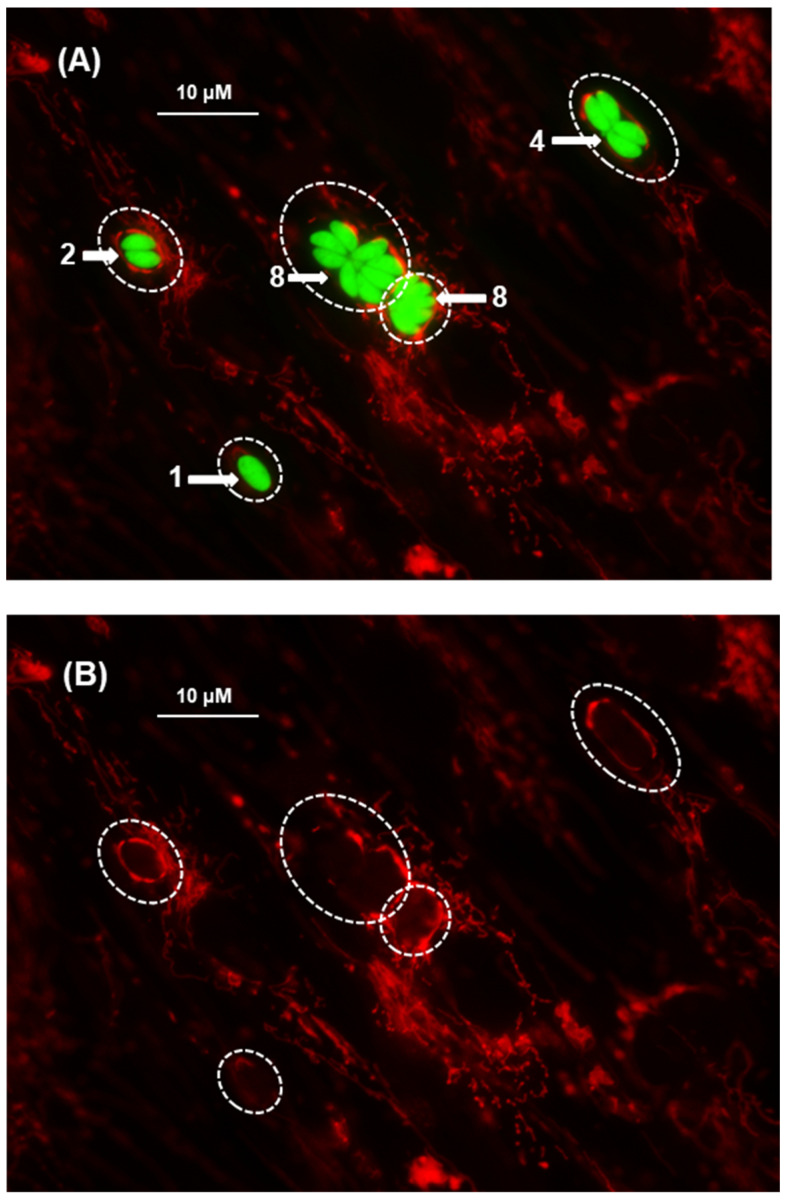
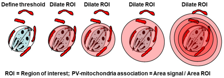
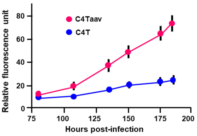
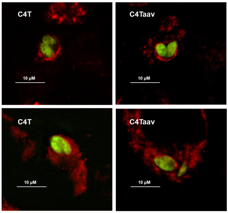
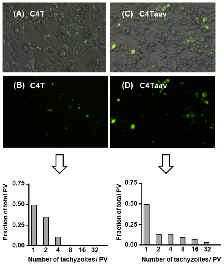
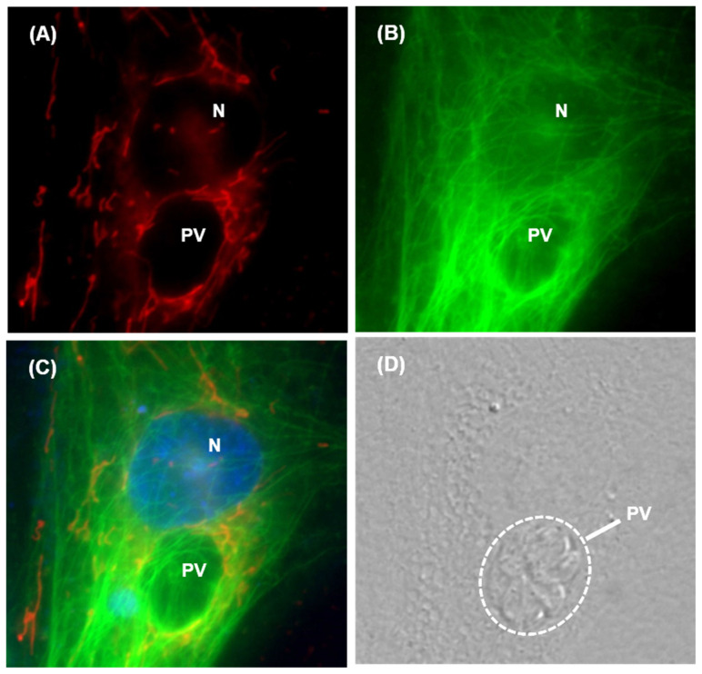
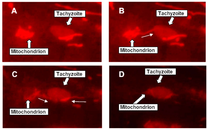
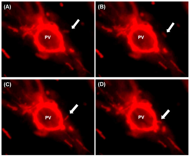
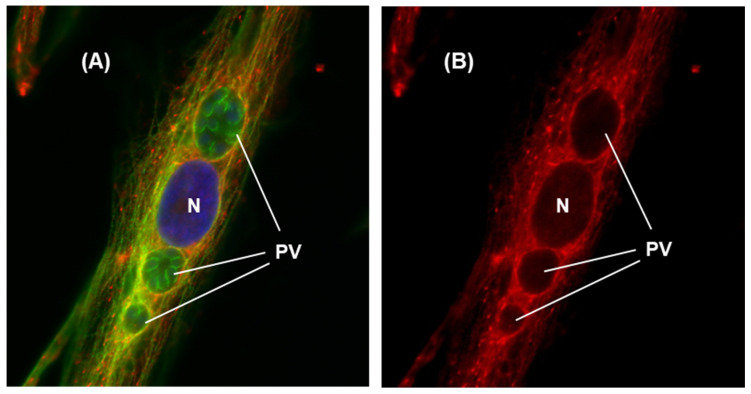
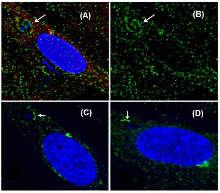
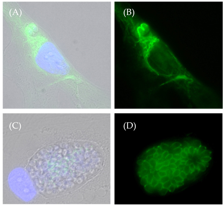
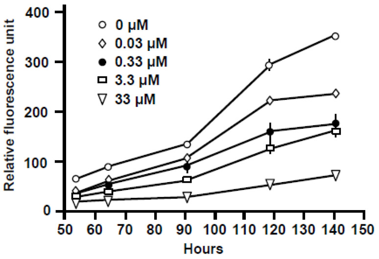
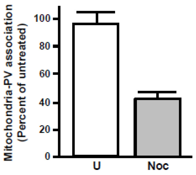
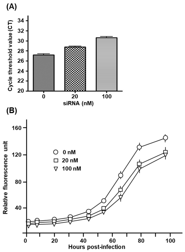
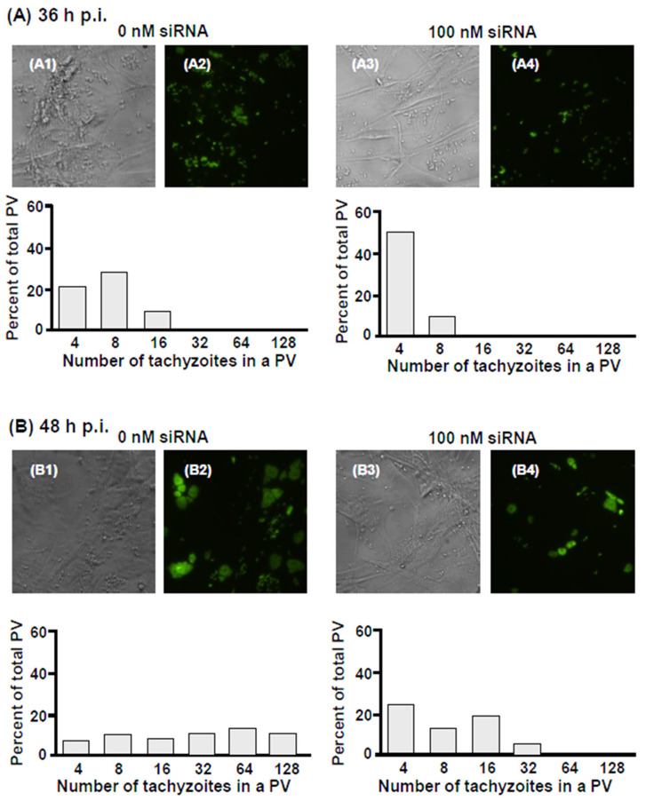
An intracellular protozoan, the Apicomplexan parasite () infects nucleated cells, in which it triggers the formation of a specialized membrane-confined cytoplasmic vacuole, named the parasitophorous vacuole (PV). One of the most prominent events in the parasite's intracellular life is the congregation of the host cell mitochondria around the PV. However, the significance of this event has remained largely unsolved since the parasite itself possesses a functional mitochondrion, which is essential for its replication. Here, we explore several fundamental aspects of the interaction between the PV and the host cell mitochondria. They include the detailed features of the congregation, the nature and mechanism of the mitochondrial travel to the PV, and the potential significance of the migration and congregation. Using a combination of biochemical assays, high-resolution imaging, and RNAi-mediated knockdown, we show that: (i) mitochondrial travel to the PV starts very early in parasite infection, as soon as the smallest PV takes shape; (ii) the travel utilizes the contractile microtubular network of the host cell; and (iii) near the end of the parasitic life cycle, when most PVs have reached their largest sustainable size and are about to lyse in order to release the progeny parasites, the associated mitochondria change their usual elongated shape to small spheres, apparently resulting from increased fission. Intriguingly, despite the well-known mitochondrial role as a major producer of cellular ATP, the parasite does not seem to use cellular mitochondrial ATP. Together, these findings may serve as foundations for future research in host-parasite interaction, particularly in the elucidation of its mechanisms, and the possible development of novel antiparasitic drug regimens.

摘要

细胞内原生动物顶复门寄生虫()感染有核细胞,在其中它会触发形成一种特殊的膜包被细胞质泡,称为寄生泡(PV)。寄生虫细胞内生命中最显著的事件之一是宿主细胞线粒体在寄生泡周围聚集。然而,由于寄生虫自身拥有一个对其复制至关重要的功能性线粒体,这一事件的意义在很大程度上仍未得到解决。在这里,我们探讨了寄生泡与宿主细胞线粒体相互作用的几个基本方面。它们包括聚集的详细特征、线粒体向寄生泡移动的性质和机制,以及迁移和聚集的潜在意义。通过结合生化分析、高分辨率成像和RNAi介导的敲低技术,我们发现:(i)线粒体向寄生泡的移动在寄生虫感染的早期就开始了,一旦最小的寄生泡形成;(ii)这种移动利用了宿主细胞的收缩微管网络;(iii)在寄生生命周期接近尾声时,当大多数寄生泡达到其最大可持续大小并即将裂解以释放子代寄生虫时,相关的线粒体将其通常的细长形状变为小球体,这显然是由于裂变增加所致。有趣的是,尽管线粒体作为细胞ATP的主要生产者的作用众所周知,但寄生虫似乎并不利用细胞线粒体产生的ATP。总之,这些发现可能为未来宿主 - 寄生虫相互作用的研究奠定基础,特别是在阐明其机制以及可能开发新型抗寄生虫药物方案方面。

https://cdn.ncbi.nlm.nih.gov/pmc/blobs/1b01/11677533/8d310df721d5/ijms-25-13459-g016.jpg
https://cdn.ncbi.nlm.nih.gov/pmc/blobs/1b01/11677533/91d383b094ed/ijms-25-13459-g001.jpg
https://cdn.ncbi.nlm.nih.gov/pmc/blobs/1b01/11677533/762690acd50b/ijms-25-13459-g002.jpg
https://cdn.ncbi.nlm.nih.gov/pmc/blobs/1b01/11677533/82c683c8c0a7/ijms-25-13459-g003.jpg
https://cdn.ncbi.nlm.nih.gov/pmc/blobs/1b01/11677533/134552d741d8/ijms-25-13459-g004.jpg
https://cdn.ncbi.nlm.nih.gov/pmc/blobs/1b01/11677533/b6a3c7d815a3/ijms-25-13459-g005.jpg
https://cdn.ncbi.nlm.nih.gov/pmc/blobs/1b01/11677533/cd0458582c92/ijms-25-13459-g006.jpg
https://cdn.ncbi.nlm.nih.gov/pmc/blobs/1b01/11677533/3c14dd35b48e/ijms-25-13459-g007.jpg
https://cdn.ncbi.nlm.nih.gov/pmc/blobs/1b01/11677533/8ea8227d5687/ijms-25-13459-g008.jpg
https://cdn.ncbi.nlm.nih.gov/pmc/blobs/1b01/11677533/afd016242f60/ijms-25-13459-g009.jpg
https://cdn.ncbi.nlm.nih.gov/pmc/blobs/1b01/11677533/3e9d01db9a93/ijms-25-13459-g010.jpg
https://cdn.ncbi.nlm.nih.gov/pmc/blobs/1b01/11677533/6a2075eec1e0/ijms-25-13459-g011.jpg
https://cdn.ncbi.nlm.nih.gov/pmc/blobs/1b01/11677533/93ce6acb7d3e/ijms-25-13459-g012.jpg
https://cdn.ncbi.nlm.nih.gov/pmc/blobs/1b01/11677533/626a33584b6a/ijms-25-13459-g013.jpg
https://cdn.ncbi.nlm.nih.gov/pmc/blobs/1b01/11677533/3e3a8bb4b2ad/ijms-25-13459-g014.jpg
https://cdn.ncbi.nlm.nih.gov/pmc/blobs/1b01/11677533/f48d2a3ccc52/ijms-25-13459-g015a.jpg
https://cdn.ncbi.nlm.nih.gov/pmc/blobs/1b01/11677533/8d310df721d5/ijms-25-13459-g016.jpg

相似文献

1
Host-Parasite Interactions in -Infected Cells: Roles of Mitochondria, Microtubules, and the Parasitophorous Vacuole.感染细胞中的宿主-寄生虫相互作用:线粒体、微管和寄生泡的作用。
Int J Mol Sci. 2024 Dec 16;25(24):13459. doi: 10.3390/ijms252413459.
2
Biogenesis of and activities at the Toxoplasma gondii parasitophorous vacuole membrane.刚地弓形虫寄生泡膜的生物发生及相关活动
Subcell Biochem. 2008;47:155-64. doi: 10.1007/978-0-387-78267-6_12.
3
Toxoplasma gondii salvages sphingolipids from the host Golgi through the rerouting of selected Rab vesicles to the parasitophorous vacuole.刚地弓形虫通过将选定的 Rab 囊泡重定向到寄生空泡,从宿主高尔基体中回收鞘脂。
Mol Biol Cell. 2013 Jun;24(12):1974-95. doi: 10.1091/mbc.E12-11-0827. Epub 2013 Apr 24.
4
association with host mitochondria requires key mitochondrial protein import machinery.与宿主线粒体的关联需要关键的线粒体蛋白输入机制。
Proc Natl Acad Sci U S A. 2021 Mar 23;118(12). doi: 10.1073/pnas.2013336118.
5
[Formation and diversity of parasitophorous vacuoles in parasitic protozoa. The Coccidia (Sporozoa, Apicomplexa)].[寄生原生动物中寄生泡的形成与多样性。球虫纲(孢子虫纲,顶复门)]
Tsitologiia. 2003;45(4):339-56.
6
GRA proteins of Toxoplasma gondii: maintenance of host-parasite interactions across the parasitophorous vacuolar membrane.刚地弓形虫的致密颗粒蛋白:维持跨寄生泡膜的宿主-寄生虫相互作用
Korean J Parasitol. 2009 Oct;47 Suppl(Suppl):S29-37. doi: 10.3347/kjp.2009.47.S.S29.
7
During host cell traversal and cell-to-cell passage, Toxoplasma gondii sporozoites inhabit the parasitophorous vacuole and posteriorly release dense granule protein-associated membranous trails.在宿主细胞穿入和细胞间传播过程中,刚地弓形虫速殖子寄生于吞噬小泡,并随后从后端释放出致密颗粒蛋白相关的膜性痕迹。
Int J Parasitol. 2020 Nov;50(13):1099-1115. doi: 10.1016/j.ijpara.2020.06.012. Epub 2020 Aug 31.
8
Association of host cell endoplasmic reticulum and mitochondria with the Toxoplasma gondii parasitophorous vacuole membrane: a high affinity interaction.宿主细胞内质网和线粒体与刚地弓形虫寄生泡膜的关联:一种高亲和力相互作用。
J Cell Sci. 1997 Sep;110 ( Pt 17):2117-28. doi: 10.1242/jcs.110.17.2117.
9
New host nuclear functions are not required for the modifications of the parasitophorous vacuole of Toxoplasma.弓形虫寄生泡的修饰不需要新的宿主核功能。
Cell Microbiol. 2008 Feb;10(2):465-76. doi: 10.1111/j.1462-5822.2007.01061.x. Epub 2007 Oct 28.
10
Molecular modifications of host cells by Toxoplasma gondii.刚地弓形虫对宿主细胞的分子修饰
Pol J Microbiol. 2004;53 Suppl:45-54.

本文引用的文献

1
Protists: Eukaryotic single-celled organisms and the functioning of their organelles.原生生物:真核单细胞生物及其细胞器的功能。
Adv Microb Physiol. 2024;84:243-307. doi: 10.1016/bs.ampbs.2024.02.001. Epub 2024 Mar 16.
2
Protein lipoylation: mitochondria, cuproptosis, and beyond.蛋白质脂酰化:线粒体、铜死亡及其他。
Trends Biochem Sci. 2024 Aug;49(8):729-744. doi: 10.1016/j.tibs.2024.04.002. Epub 2024 May 6.
3
Contact sites between host organelles and pathogens: boon or bane?宿主细胞器与病原体的接触部位:是福还是祸?
mSphere. 2023 Dec 20;8(6):e0044823. doi: 10.1128/msphere.00448-23. Epub 2023 Oct 18.
4
High burden and low awareness of toxoplasmosis in the United States.美国弓形虫病负担高而知晓率低。
Postgrad Med. 2019 Mar;131(2):103-108. doi: 10.1080/00325481.2019.1568792. Epub 2019 Jan 21.
5
Association of mitochondria with microtubules inhibits mitochondrial fission by precluding assembly of the fission protein Dnm1.线粒体与微管的结合通过阻止分裂蛋白 Dnm1 的组装来抑制线粒体的分裂。
J Biol Chem. 2019 Mar 8;294(10):3385-3396. doi: 10.1074/jbc.RA118.006799. Epub 2019 Jan 2.
6
A Toxoplasma gondii locus required for the direct manipulation of host mitochondria has maintained multiple ancestral functions.刚地弓形虫中一个直接操纵宿主线粒体必需的基因座,保持了多种祖先功能。
Mol Microbiol. 2018 Jun;108(5):519-535. doi: 10.1111/mmi.13947. Epub 2018 Apr 11.
7
Protein lipoylation: an evolutionarily conserved metabolic regulator of health and disease.蛋白质脂酰化:一种进化上保守的代谢调节剂,影响健康和疾病。
Curr Opin Chem Biol. 2018 Feb;42:76-85. doi: 10.1016/j.cbpa.2017.11.003. Epub 2017 Nov 21.
8
Identification of compounds that suppress Toxoplasma gondii tachyzoites and bradyzoites.鉴定抑制刚地弓形虫速殖子和缓殖子的化合物。
PLoS One. 2017 Jun 13;12(6):e0178203. doi: 10.1371/journal.pone.0178203. eCollection 2017.
9
Host Mitochondrial Association Evolved in the Human Parasite Toxoplasma gondii via Neofunctionalization of a Gene Duplicate.宿主线粒体关联通过基因复制的新功能化在人类寄生虫弓形虫中进化而来。
Genetics. 2016 May;203(1):283-98. doi: 10.1534/genetics.115.186270. Epub 2016 Feb 26.
10
Neurodegeneration and microtubule dynamics: death by a thousand cuts.神经退行性变与微管动力学:千刀万剐式死亡
Front Cell Neurosci. 2015 Sep 9;9:343. doi: 10.3389/fncel.2015.00343. eCollection 2015.