Jiang Yinjie, Chen Xun, Cheng Mingrui, Lin I-Chun, Li Boliang, Zhu Xinjie, Xu Guanghan, Miao Huamao, Wang Xiaoying, Zhou Xingtao
Fudan University Eye Ear Nose and Throat Hospital, Shanghai, China.
Key laboratory of Myopia and Related Eye Diseases, National Health Commission, Shanghai, China.
BMC Ophthalmol. 2025 Jan 7;25(1):10. doi: 10.1186/s12886-024-03803-0.
To evaluate the biosafety, reduction in anterior capsule opacification, and fluctuation in intraocular pressure (IOP) of a new phakic refractive lens (PRL) with a sinusoidal drainage groove design.
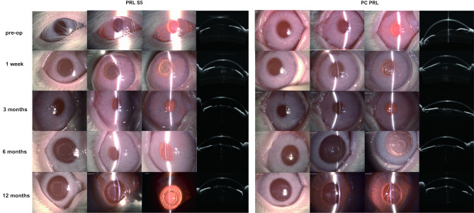
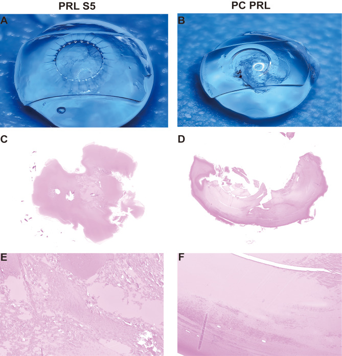
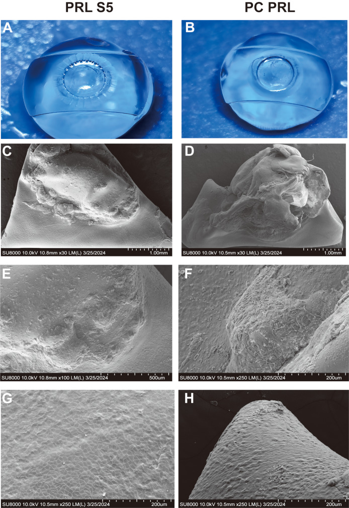
This self-controlled experiment was performed on eight eyes of four rabbits. Each rabbit was implanted with a sinusoidal PRL (PRL-S5) in the right eye and a conventional posterior chamber PRL (PC-PRL) in the left eye. Slit-lamp examinations, optical coherence tomography, and IOP evaluation were performed before surgery and at 1 day, 1 week, 3 months, 6 months, and 1 year postoperatively in each eye. Gross examination, histopathology, and electron microscopy of the capsule and PRL were performed 1 year postoperatively.
On slit-lamp examination, the inflammatory reactions recovered one week after surgery. The PC-PRL group developed anterior subcapsular cataracts at 3 months postoperatively and diffuse and dense opacification of the cortex at 1 year. PRL-S5 showed mild local opacification in the optical zone 6 months postoperatively, which did not progress significantly. At 1 year, PC-PRLs had greater opacification (27.37-72.17%) than PRL S5 (6.63-66.96%). Three months after surgery, one eye in the PC-PRL group had scleral staphyloma, one eye had corneal edema, and one eye experienced nasal hepatic prolapse into the anterior chamber. One eye in the PRL-S5 group had papillary membranes but recovered 6 months postoperatively. Histopathological examination revealed liquefaction and necrosis of the opacified area in the center of the subcapsular membrane in both groups. Numerous granular bodies and fibrous precipitates were observed in epithelial cells in the opaque area. Electron microscopy showed that epithelial cells proliferated on the surface of all anterior capsule membranes, with no significant differences between the two groups. The capsular PC-PRL group showed anterior cortical proliferation and fibrosis. An IOP elevation was noted on the first postoperative day (18.8 to 37.9 mmHg). However, both the PC-PRL and PRL-S5 groups exhibited relatively stable IOP levels 1 week, 3 months, 6 months, and 1 year postoperatively.
The new PRL exhibited robust long-term biocompatibility. The sinusoidal groove design facilitated the maintenance of IOP stability without necessitating iridectomy and effectively mitigated the onset and progression of cataracts.
评估一种具有正弦引流槽设计的新型有晶状体眼屈光晶状体(PRL)的生物安全性、前囊膜混浊程度的降低情况以及眼内压(IOP)的波动。
本自我对照实验在4只兔子的8只眼睛上进行。每只兔子右眼植入正弦PRL(PRL-S5),左眼植入传统后房型PRL(PC-PRL)。在手术前以及术后第1天、1周、3个月、6个月和1年对每只眼睛进行裂隙灯检查、光学相干断层扫描和IOP评估。术后1年对晶状体囊膜和PRL进行大体检查、组织病理学检查和电子显微镜检查。
裂隙灯检查显示,术后1周炎症反应消退。PC-PRL组术后3个月出现前囊下白内障,术后1年皮质出现弥漫性致密混浊。PRL-S5术后6个月在光学区出现轻度局部混浊,且未显著进展。术后1年,PC-PRL的混浊程度(27.37%-72.17%)高于PRL-S5(6.63%-66.96%)。术后3个月,PC-PRL组1只眼睛出现巩膜葡萄肿,1只眼睛出现角膜水肿,1只眼睛出现鼻侧虹膜脱垂至前房。PRL-S5组1只眼睛出现瞳孔膜,但术后6个月恢复。组织病理学检查显示两组晶状体囊膜下中央混浊区域均有液化和坏死。在混浊区域的上皮细胞中观察到大量颗粒体和纤维沉淀物。电子显微镜检查显示所有前囊膜表面上皮细胞均有增殖,两组之间无显著差异。PC-PRL组晶状体囊膜显示前皮质增殖和纤维化。术后第1天IOP升高(18.8至37.9 mmHg)。然而,PC-PRL组和PRL-S5组术后1周、3个月、6个月和1年的IOP水平均相对稳定。
新型PRL具有良好的长期生物相容性。正弦槽设计有助于维持IOP稳定,无需行虹膜切除术,并有效减轻白内障的发生和进展。