• 文献检索
  • 文档翻译
  • 深度研究
  • 学术资讯
  • Suppr Zotero 插件Zotero 插件
  • 邀请有礼
  • 套餐&价格
  • 历史记录
应用&插件
Suppr Zotero 插件Zotero 插件浏览器插件Mac 客户端Windows 客户端微信小程序
定价
高级版会员购买积分包购买API积分包
服务
文献检索文档翻译深度研究API 文档MCP 服务
关于我们
关于 Suppr公司介绍联系我们用户协议隐私条款
关注我们

Suppr 超能文献

核心技术专利:CN118964589B侵权必究
粤ICP备2023148730 号-1Suppr @ 2026

文献检索

告别复杂PubMed语法,用中文像聊天一样搜索,搜遍4000万医学文献。AI智能推荐,让科研检索更轻松。

立即免费搜索

文件翻译

保留排版,准确专业,支持PDF/Word/PPT等文件格式,支持 12+语言互译。

免费翻译文档

深度研究

AI帮你快速写综述,25分钟生成高质量综述,智能提取关键信息,辅助科研写作。

立即免费体验

胎儿肝脏造血微环境的时空动态变化

Spatiotemporal dynamics of fetal liver hematopoietic niches.

作者信息

Mesquita Peixoto Márcia, Soares-da-Silva Francisca, Bonnet Valentin, Zhou Yanping, Ronteix Gustave, Santos Rita Faria, Mailhe Marie-Pierre, Nogueira Gonçalo, Feng Xing, Pereira João Pedro, Azzoni Emanuele, Anselmi Giorgio, de Bruijn Marella F T R, Perkins Archibald, Baroud Charles N, Pinto-do-Ó Perpétua, Cumano Ana

机构信息

Immunology Department, Unit of Lymphocytes and Immunity, Institut Pasteur, Paris, France.

INSERM U1223 , Paris, France.

出版信息

J Exp Med. 2025 Feb 3;222(2). doi: 10.1084/jem.20240592. Epub 2025 Jan 7.

DOI:10.1084/jem.20240592
PMID:39775824
原文链接:https://pmc.ncbi.nlm.nih.gov/articles/PMC11706214/
Abstract

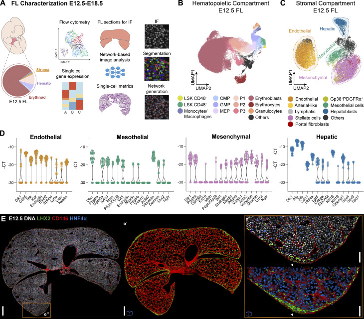
Embryonic hematopoietic cells develop in the fetal liver (FL), surrounded by diverse non-hematopoietic stromal cells. However, the spatial organization and cytokine production patterns of the stroma during FL development remain poorly understood. Here, we characterized and mapped the hematopoietic and stromal cell populations at early (E12.5-14.5) FL stages, revealing that while hepatoblasts were the primary source of hematopoietic growth factors, other stromal cells-including mesenchymal, mesothelial, and endothelial cells-also contributed to this signaling network. Using a dedicated image analysis pipeline, we quantified cell distances to tissue structures and defined neighbor relationships, uncovering that different hematopoietic progenitors exhibit distinct preferences for neighboring stromal cells and show developmental changes in spatial distribution. Notably, our data suggest that the sub-mesothelium region plays a prominent role in early fetal hematopoiesis. This approach offers a valuable tool for studying complex cellular interactions in biological systems, providing new insights into hematopoietic niche organization during development.

摘要

胚胎造血细胞在胎儿肝脏(FL)中发育,周围是各种非造血基质细胞。然而,在FL发育过程中基质的空间组织和细胞因子产生模式仍知之甚少。在这里,我们对早期(E12.5 - 14.5)FL阶段的造血和基质细胞群体进行了表征和绘图,发现虽然肝细胞是造血生长因子的主要来源,但其他基质细胞,包括间充质细胞、间皮细胞和内皮细胞,也参与了这个信号网络。使用专门的图像分析流程,我们量化了细胞与组织结构的距离并定义了邻域关系,发现不同的造血祖细胞对相邻基质细胞表现出不同的偏好,并在空间分布上显示出发育变化。值得注意的是,我们的数据表明间皮下区域在早期胎儿造血中起重要作用。这种方法为研究生物系统中复杂的细胞相互作用提供了有价值的工具,为发育过程中的造血微环境组织提供了新的见解。

https://cdn.ncbi.nlm.nih.gov/pmc/blobs/f390/11706214/ee5a781ab0eb/jem_20240592_fig9.jpg
https://cdn.ncbi.nlm.nih.gov/pmc/blobs/f390/11706214/cc6a342c20b8/jem_20240592_fig1.jpg
https://cdn.ncbi.nlm.nih.gov/pmc/blobs/f390/11706214/ca2502f633bf/jem_20240592_figs1.jpg
https://cdn.ncbi.nlm.nih.gov/pmc/blobs/f390/11706214/5739cbf68a88/jem_20240592_fig2.jpg
https://cdn.ncbi.nlm.nih.gov/pmc/blobs/f390/11706214/fadfe540ea37/jem_20240592_fig3.jpg
https://cdn.ncbi.nlm.nih.gov/pmc/blobs/f390/11706214/3cfe1dded747/jem_20240592_figs2.jpg
https://cdn.ncbi.nlm.nih.gov/pmc/blobs/f390/11706214/f0fb91a383cc/jem_20240592_fig4.jpg
https://cdn.ncbi.nlm.nih.gov/pmc/blobs/f390/11706214/60af0e04244d/jem_20240592_figs3.jpg
https://cdn.ncbi.nlm.nih.gov/pmc/blobs/f390/11706214/7de497f99761/jem_20240592_fig5.jpg
https://cdn.ncbi.nlm.nih.gov/pmc/blobs/f390/11706214/e3d9e3b63037/jem_20240592_fig6.jpg
https://cdn.ncbi.nlm.nih.gov/pmc/blobs/f390/11706214/f5568d95eac1/jem_20240592_figs4.jpg
https://cdn.ncbi.nlm.nih.gov/pmc/blobs/f390/11706214/242104588dcd/jem_20240592_fig7.jpg
https://cdn.ncbi.nlm.nih.gov/pmc/blobs/f390/11706214/3b7a490ce9ca/jem_20240592_figs5.jpg
https://cdn.ncbi.nlm.nih.gov/pmc/blobs/f390/11706214/532d9ee83f39/jem_20240592_fig8.jpg
https://cdn.ncbi.nlm.nih.gov/pmc/blobs/f390/11706214/ee5a781ab0eb/jem_20240592_fig9.jpg

相似文献

1
Spatiotemporal dynamics of fetal liver hematopoietic niches.胎儿肝脏造血微环境的时空动态变化
J Exp Med. 2025 Feb 3;222(2). doi: 10.1084/jem.20240592. Epub 2025 Jan 7.
2
Clonal precursor of bone, cartilage, and hematopoietic niche stromal cells.骨、软骨和造血龛基质细胞的克隆前体细胞。
Proc Natl Acad Sci U S A. 2013 Jul 30;110(31):12643-8. doi: 10.1073/pnas.1310212110. Epub 2013 Jul 15.
3
Molecular signature and in vivo behavior of bone marrow endosteal and subendosteal stromal cell populations and their relevance to hematopoiesis.骨髓骨内膜和骨内膜下基质细胞群体的分子特征和体内行为及其与造血的关系。
Exp Cell Res. 2012 Nov 15;318(19):2427-37. doi: 10.1016/j.yexcr.2012.07.009. Epub 2012 Jul 27.
4
[Expansion of bone marrow LTC-ICs in vitro by mouse fetal liver-derived stromal cell lines].[小鼠胎肝来源的基质细胞系在体外对骨髓长期培养起始细胞的扩增作用]
Sheng Wu Gong Cheng Xue Bao. 2003 Jul;19(4):450-5.
5
Fetal stromal niches enhance human embryonic stem cell-derived hematopoietic differentiation and globin switch.胎儿基质龛增强人胚胎干细胞来源的造血分化和珠蛋白开关。
Stem Cells Dev. 2011 Jan;20(1):31-8. doi: 10.1089/scd.2010.0196. Epub 2010 Oct 12.
6
ATF4 plays a pivotal role in the development of functional hematopoietic stem cells in mouse fetal liver.激活转录因子4(ATF4)在小鼠胎肝中功能性造血干细胞的发育过程中起着关键作用。
Blood. 2015 Nov 19;126(21):2383-91. doi: 10.1182/blood-2015-03-633354. Epub 2015 Sep 17.
7
Hematopoietic supportive functions of mouse bone marrow and fetal liver microenvironment: dissection of granulocyte, B-lymphocyte, and hematopoietic progenitor support at the stroma cell clone level.小鼠骨髓和胎肝微环境的造血支持功能:在基质细胞克隆水平剖析粒细胞、B淋巴细胞和造血祖细胞的支持作用。
Blood. 1996 Jun 1;87(11):4596-606.
8
Spatiotemporal dynamics of cytokines expression dictate fetal liver hematopoiesis.细胞因子表达的时空动态决定了胎儿肝脏造血。
bioRxiv. 2023 Aug 25:2023.08.24.554612. doi: 10.1101/2023.08.24.554612.
9
Towards Mimicking the Fetal Liver Niche: The Influence of Elasticity and Oxygen Tension on Hematopoietic Stem/Progenitor Cells Cultured in 3D Fibrin Hydrogels.迈向模拟胎儿肝脏微环境:弹性和氧张力对三维纤维蛋白水凝胶中培养的造血干/祖细胞的影响。
Int J Mol Sci. 2020 Sep 2;21(17):6367. doi: 10.3390/ijms21176367.
10
Analysis of the human fetal liver hematopoietic microenvironment.人类胎儿肝脏造血微环境分析
Stem Cells Dev. 2005 Oct;14(5):493-504. doi: 10.1089/scd.2005.14.493.

引用本文的文献

1
A Neutrophil Storage Pool in the Fetal Liver is the Principal Source of the Postnatal Neutrophil Surge.胎儿肝脏中的中性粒细胞储存池是出生后中性粒细胞激增的主要来源。
FASEB J. 2025 Aug 15;39(15):e70921. doi: 10.1096/fj.202501613RR.
2
Resolving the spatial organization of fetal liver hematopoiesis by SeekSpace.利用SeekSpace解析胎儿肝脏造血的空间组织。
Cell Regen. 2025 Apr 22;14(1):15. doi: 10.1186/s13619-025-00234-0.

本文引用的文献

1
Assembling the layers of the hematopoietic system: A window of opportunity for thymopoiesis in the embryo.组装造血系统的层次结构:胚胎中胸腺发生的机会之窗。
Immunol Rev. 2023 May;315(1):54-70. doi: 10.1111/imr.13187. Epub 2023 Mar 3.
2
Murine foetal liver supports limited detectable expansion of life-long haematopoietic progenitors.鼠胎肝支持有限的可检测到的终生造血祖细胞的扩增。
Nat Cell Biol. 2022 Oct;24(10):1475-1486. doi: 10.1038/s41556-022-00999-5. Epub 2022 Oct 6.
3
Independent origins of fetal liver haematopoietic stem and progenitor cells.
胎儿肝脏造血干细胞和祖细胞的独立起源。
Nature. 2022 Sep;609(7928):779-784. doi: 10.1038/s41586-022-05203-0. Epub 2022 Sep 14.
4
Griottes: a generalist tool for network generation from segmented tissue images.Griottes:一种从分割组织图像生成网络的通用工具。
BMC Biol. 2022 Aug 11;20(1):178. doi: 10.1186/s12915-022-01376-2.
5
: Development, Genetic and Epigenetic Abnormalities.发育、遗传和表观遗传异常。
Cells. 2022 Jun 10;11(12):1886. doi: 10.3390/cells11121886.
6
Identification of fetal liver stroma in spectral cytometry using the parameter autofluorescence.使用参数自发荧光对光谱细胞术中小鼠胎肝基质细胞的鉴定。
Cytometry A. 2022 Nov;101(11):960-969. doi: 10.1002/cyto.a.24567. Epub 2022 May 14.
7
Hematopoietic stem cells temporally transition to thrombopoietin dependence in the fetal liver.造血干细胞在胎肝中暂时过渡到对血小板生成素的依赖。
Sci Adv. 2022 Mar 18;8(11):eabm7688. doi: 10.1126/sciadv.abm7688. Epub 2022 Mar 16.
8
Mds1, an inducible Cre allele specific to adult-repopulating hematopoietic stem cells.Mds1,一种诱导型 Cre 等位基因,特异性针对成人造血干细胞。
Cell Rep. 2021 Aug 17;36(7):109562. doi: 10.1016/j.celrep.2021.109562.
9
Yolk sac, but not hematopoietic stem cell-derived progenitors, sustain erythropoiesis throughout murine embryonic life.卵黄囊,但不是造血干细胞衍生的祖细胞,在整个小鼠胚胎生命过程中维持红细胞生成。
J Exp Med. 2021 Apr 5;218(4). doi: 10.1084/jem.20201729.
10
Hematopoiesis: A Layered Organization Across Chordate Species.造血作用:脊索动物物种中的分层组织
Front Cell Dev Biol. 2020 Dec 16;8:606642. doi: 10.3389/fcell.2020.606642. eCollection 2020.