• 文献检索
  • 文档翻译
  • 深度研究
  • 学术资讯
  • Suppr Zotero 插件Zotero 插件
  • 邀请有礼
  • 套餐&价格
  • 历史记录
应用&插件
Suppr Zotero 插件Zotero 插件浏览器插件Mac 客户端Windows 客户端微信小程序
定价
高级版会员购买积分包购买API积分包
服务
文献检索文档翻译深度研究API 文档MCP 服务
关于我们
关于 Suppr公司介绍联系我们用户协议隐私条款
关注我们

Suppr 超能文献

核心技术专利:CN118964589B侵权必究
粤ICP备2023148730 号-1Suppr @ 2026

文献检索

告别复杂PubMed语法,用中文像聊天一样搜索,搜遍4000万医学文献。AI智能推荐,让科研检索更轻松。

立即免费搜索

文件翻译

保留排版,准确专业,支持PDF/Word/PPT等文件格式,支持 12+语言互译。

免费翻译文档

深度研究

AI帮你快速写综述,25分钟生成高质量综述,智能提取关键信息,辅助科研写作。

立即免费体验

Griottes:一种从分割组织图像生成网络的通用工具。

Griottes: a generalist tool for network generation from segmented tissue images.

机构信息

Institut Pasteur, Université Paris Cité, Physical microfluidics and Bioengineering, Paris, F-75015, France.

LadHyX, CNRS, Ecole Polytechnique, Institut Polytechnique de Paris, Palaiseau, 91120, France.

出版信息

BMC Biol. 2022 Aug 11;20(1):178. doi: 10.1186/s12915-022-01376-2.

DOI:10.1186/s12915-022-01376-2
PMID:35953853
原文链接:https://pmc.ncbi.nlm.nih.gov/articles/PMC9367069/
Abstract

BACKGROUND

Microscopy techniques and image segmentation algorithms have improved dramatically this decade, leading to an ever increasing amount of biological images and a greater reliance on imaging to investigate biological questions. This has created a need for methods to extract the relevant information on the behaviors of cells and their interactions, while reducing the amount of computing power required to organize this information.

RESULTS

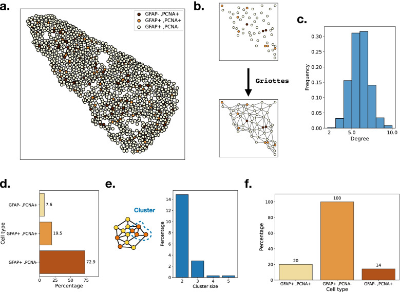
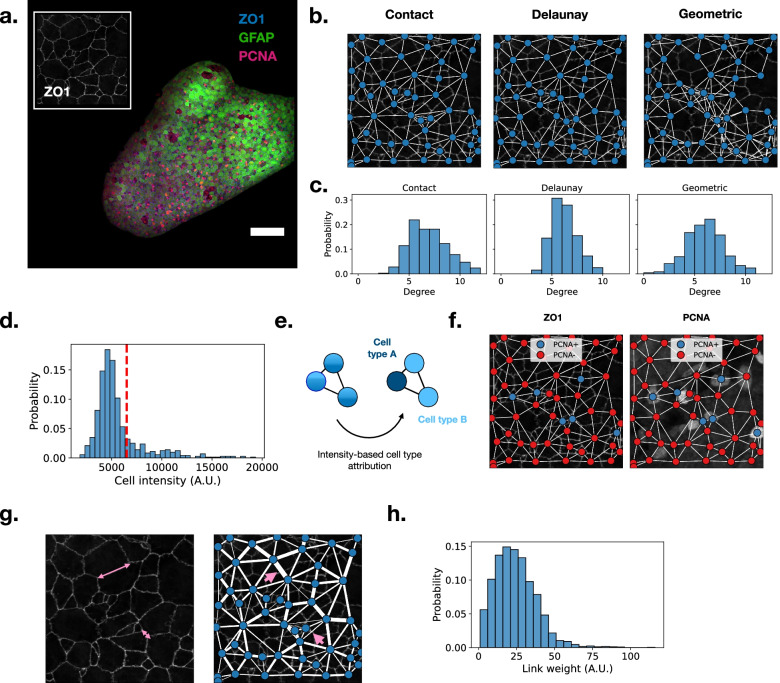
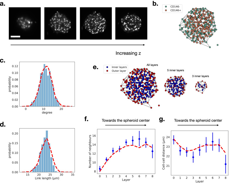
This task can be performed by using a network representation in which the cells and their properties are encoded in the nodes, while the neighborhood interactions are encoded by the links. Here, we introduce Griottes, an open-source tool to build the "network twin" of 2D and 3D tissues from segmented microscopy images. We show how the library can provide a wide range of biologically relevant metrics on individual cells and their neighborhoods, with the objective of providing multi-scale biological insights. The library's capacities are demonstrated on different image and data types.

CONCLUSIONS

This library is provided as an open-source tool that can be integrated into common image analysis workflows to increase their capacities.

摘要

背景

显微镜技术和图像分割算法在过去十年中得到了显著的改进,导致生物图像的数量不断增加,并且对成像技术的依赖程度也越来越高,以研究生物学问题。这就需要开发方法来提取有关细胞行为及其相互作用的相关信息,同时减少组织这些信息所需的计算能力。

结果

可以通过使用网络表示来执行此任务,其中将细胞及其属性编码为节点,而将邻域交互编码为链接。在这里,我们介绍了 Griottes,这是一个开源工具,用于从分割的显微镜图像构建 2D 和 3D 组织的“网络孪生体”。我们展示了该库如何为单个细胞及其邻居提供广泛的生物学相关指标,旨在提供多尺度的生物学见解。该库的功能已在不同的图像和数据类型上得到了验证。

结论

该库作为一个开源工具提供,可以集成到常见的图像分析工作流程中,以提高其能力。

https://cdn.ncbi.nlm.nih.gov/pmc/blobs/05a0/9367069/4682bb18f807/12915_2022_1376_Fig4_HTML.jpg
https://cdn.ncbi.nlm.nih.gov/pmc/blobs/05a0/9367069/7980346724e4/12915_2022_1376_Fig1_HTML.jpg
https://cdn.ncbi.nlm.nih.gov/pmc/blobs/05a0/9367069/942806b12d42/12915_2022_1376_Fig2_HTML.jpg
https://cdn.ncbi.nlm.nih.gov/pmc/blobs/05a0/9367069/03bea46daae5/12915_2022_1376_Fig3_HTML.jpg
https://cdn.ncbi.nlm.nih.gov/pmc/blobs/05a0/9367069/4682bb18f807/12915_2022_1376_Fig4_HTML.jpg

相似文献

1
Griottes: a generalist tool for network generation from segmented tissue images.Griottes:一种从分割组织图像生成网络的通用工具。
BMC Biol. 2022 Aug 11;20(1):178. doi: 10.1186/s12915-022-01376-2.
2
Polymorph segmentation representation for medical image computing.多态分割表示在医学图像计算中的应用。
Comput Methods Programs Biomed. 2019 Apr;171:19-26. doi: 10.1016/j.cmpb.2019.02.011. Epub 2019 Feb 21.
3
Svetlana a supervised segmentation classifier for Napari.斯维特拉娜监督了 Napari 的分割分类器。
Sci Rep. 2024 May 21;14(1):11604. doi: 10.1038/s41598-024-60916-8.
4
PartSeg: a tool for quantitative feature extraction from 3D microscopy images for dummies.PartSeg:傻瓜式 3D 显微镜图像定量特征提取工具。
BMC Bioinformatics. 2021 Feb 17;22(1):72. doi: 10.1186/s12859-021-03984-1.
5
Femoral image segmentation based on two-stage convolutional network using 3D-DMFNet and 3D-ResUnet.基于使用 3D-DMFNet 和 3D-ResUnet 的两阶段卷积网络的股骨图像分割。
Comput Methods Programs Biomed. 2022 Nov;226:107110. doi: 10.1016/j.cmpb.2022.107110. Epub 2022 Sep 6.
6
Metrics for evaluating 3D medical image segmentation: analysis, selection, and tool.用于评估3D医学图像分割的指标:分析、选择与工具
BMC Med Imaging. 2015 Aug 12;15:29. doi: 10.1186/s12880-015-0068-x.
7
CellWalker: a user-friendly and modular computational pipeline for morphological analysis of microscopy images.CellWalker:一个用于显微镜图像形态分析的用户友好且模块化的计算流程。
Bioinformatics. 2023 Dec 1;39(12). doi: 10.1093/bioinformatics/btad710.
8
Fast plaque burden assessment of the femoral artery using 3D black-blood MRI and automated segmentation.使用 3D 黑血 MRI 和自动分割技术快速评估股动脉斑块负担。
Med Phys. 2011 Oct;38(10):5370-84. doi: 10.1118/1.3633899.
9
Bi-channel image registration and deep-learning segmentation (BIRDS) for efficient, versatile 3D mapping of mouse brain.双通道图像配准和深度学习分割(BIRDS)用于高效、通用的小鼠大脑 3D 映射。
Elife. 2021 Jan 18;10:e63455. doi: 10.7554/eLife.63455.
10
EmbedSeg: Embedding-based Instance Segmentation for Biomedical Microscopy Data.EmbedSeg:基于嵌入的生物医学显微镜数据实例分割。
Med Image Anal. 2022 Oct;81:102523. doi: 10.1016/j.media.2022.102523. Epub 2022 Jul 3.

引用本文的文献

1
CellMet: Extracting 3D shape and topology metrics from confluent cells within tissues.CellMet:从组织内融合细胞中提取三维形状和拓扑度量。
PLoS Comput Biol. 2025 Jul 30;21(7):e1013260. doi: 10.1371/journal.pcbi.1013260. eCollection 2025 Jul.
2
CD103+CD56+ ILCs Are Associated with an Altered CD8+ T-cell Profile within the Tumor Microenvironment.CD103+CD56+固有淋巴细胞与肿瘤微环境中CD8+ T细胞谱的改变有关。
Cancer Immunol Res. 2025 Apr 2;13(4):527-546. doi: 10.1158/2326-6066.CIR-24-0151.
3
Polarity-JaM: an image analysis toolbox for cell polarity, junction and morphology quantification.

本文引用的文献

1
Tysserand-fast and accurate reconstruction of spatial networks from bioimages.Tysserand——从生物图像中快速准确地重建空间网络。
Bioinformatics. 2021 Nov 5;37(21):3989-3991. doi: 10.1093/bioinformatics/btab490.
2
Characterizing spatial gene expression heterogeneity in spatially resolved single-cell transcriptomic data with nonuniform cellular densities.刻画具有非均匀细胞密度的空间分辨单细胞转录组数据中空间基因表达异质性。
Genome Res. 2021 Oct;31(10):1843-1855. doi: 10.1101/gr.271288.120. Epub 2021 May 25.
3
Dynamic spatiotemporal coordination of neural stem cell fate decisions occurs through local feedback in the adult vertebrate brain.
极性-JaM:用于细胞极性、连接和形态定量分析的图像分析工具箱。
Nat Commun. 2025 Feb 8;16(1):1474. doi: 10.1038/s41467-025-56643-x.
4
Spatiotemporal dynamics of fetal liver hematopoietic niches.胎儿肝脏造血微环境的时空动态变化
J Exp Med. 2025 Feb 3;222(2). doi: 10.1084/jem.20240592. Epub 2025 Jan 7.
5
Homebuilt Imaging-Based Spatial Transcriptomics: Tertiary Lymphoid Structures as a Case Example.基于自建成像的空间转录组学:以三级淋巴结构为例。
Methods Mol Biol. 2025;2864:77-105. doi: 10.1007/978-1-0716-4184-2_5.
6
Tissues as networks of cells: towards generative rules of complex organ development.组织作为细胞网络:复杂器官发育的生成规则。
J R Soc Interface. 2023 Jul;20(204):20230115. doi: 10.1098/rsif.2023.0115. Epub 2023 Jul 26.
7
The promise and challenge of spatial omics in dissecting tumour microenvironment and the role of AI.空间组学在剖析肿瘤微环境中的前景与挑战以及人工智能的作用
Front Oncol. 2023 May 1;13:1172314. doi: 10.3389/fonc.2023.1172314. eCollection 2023.
神经干细胞命运决策的动态时空协调是通过成年脊椎动物大脑中的局部反馈来实现的。
Cell Stem Cell. 2021 Aug 5;28(8):1457-1472.e12. doi: 10.1016/j.stem.2021.03.014. Epub 2021 Apr 5.
4
Cellpose: a generalist algorithm for cellular segmentation.Cellpose:一种通用的细胞分割算法。
Nat Methods. 2021 Jan;18(1):100-106. doi: 10.1038/s41592-020-01018-x. Epub 2020 Dec 14.
5
High-Throughput Measurements of Intra-Cellular and Secreted Cytokine from Single Spheroids Using Anchored Microfluidic Droplets.使用锚定微流控液滴对单个球体中的细胞内和分泌细胞因子进行高通量测量。
Small. 2020 Dec;16(49):e2002303. doi: 10.1002/smll.202002303. Epub 2020 Nov 13.
6
Coordinated Cellular Neighborhoods Orchestrate Antitumoral Immunity at the Colorectal Cancer Invasive Front.协调细胞邻里关系在结直肠癌侵袭前沿调控抗肿瘤免疫。
Cell. 2020 Sep 3;182(5):1341-1359.e19. doi: 10.1016/j.cell.2020.07.005. Epub 2020 Aug 6.
7
Individual Control and Quantification of 3D Spheroids in a High-Density Microfluidic Droplet Array.在高密度微流控液滴阵列中对 3D 球体进行个体控制和量化。
Cell Rep. 2020 May 26;31(8):107670. doi: 10.1016/j.celrep.2020.107670.
8
CytoMAP: A Spatial Analysis Toolbox Reveals Features of Myeloid Cell Organization in Lymphoid Tissues.CytoMAP:揭示淋巴组织中髓样细胞组织特征的空间分析工具箱。
Cell Rep. 2020 Apr 21;31(3):107523. doi: 10.1016/j.celrep.2020.107523.
9
Mapping the structure and biological functions within mesenchymal bodies using microfluidics.利用微流控技术绘制间充质体的结构和生物学功能图谱。
Sci Adv. 2020 Mar 4;6(10):eaaw7853. doi: 10.1126/sciadv.aaw7853. eCollection 2020 Mar.
10
The single-cell pathology landscape of breast cancer.乳腺癌的单细胞病理学图谱。
Nature. 2020 Feb;578(7796):615-620. doi: 10.1038/s41586-019-1876-x. Epub 2020 Jan 20.