Sapp Ellen, Boudi Adel, Iwanowicz Andrew, Belgrad Jillian, Miller Rachael, O'Reilly Daniel, Yamada Ken, Deng Yunping, Joni Marion, Li Xueyi, Kegel-Gleason Kimberly, Khvorova Anastasia, Reiner Anton, Aronin Neil, DiFiglia Marian
Department of Neurology, Massachusetts General Hospital, Charlestown, MA 02129.
RNA Therapeutics Institute, University of Massachusetts Chan Medical School, Worcester, MA 01605.
bioRxiv. 2025 Jan 1:2024.12.31.630891. doi: 10.1101/2024.12.31.630891.
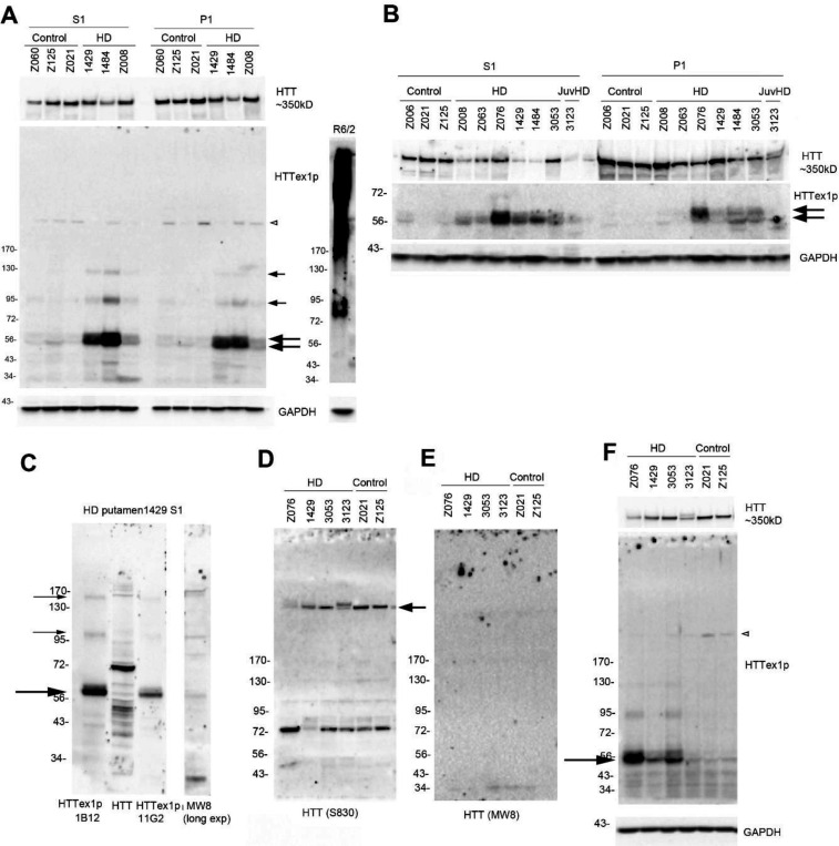
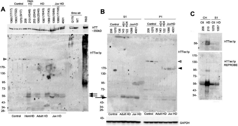
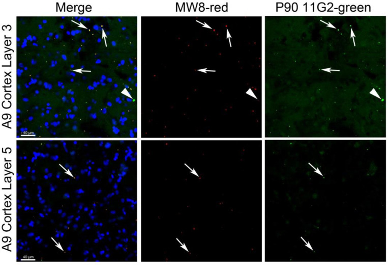
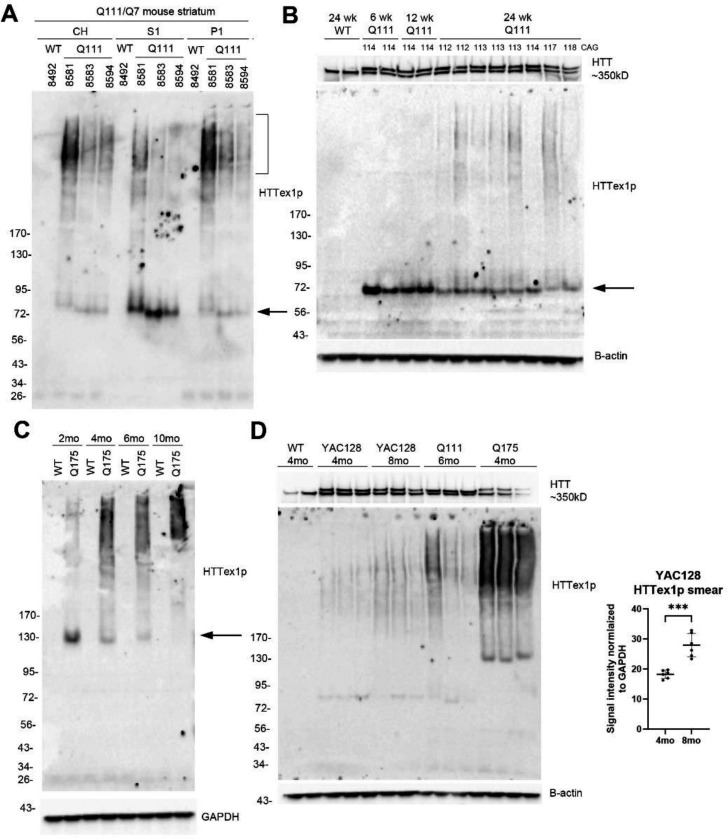
has been identified in human and mouse HD brain as the pathogenic exon 1 mRNA generated from aberrant splicing between exon 1 and 2 that contributes to aggregate formation and neuronal dysfunction (Sathasivam et al., 2013). Detection of the HTT exon 1 protein (HTTex1p) has been accomplished with surrogate antibodies in fluorescence-based reporter assays (MSD, HTRF), and immunoprecipitation assays, in HD postmortem cerebellum and knock-in mice but direct detection by SDS-PAGE and western blot assay has been lacking. Here proteins in subcellular fractions prepared from human and mouse HD brain were separated by SDS-PAGE and probed by western blot with neo-epitope monoclonal antibodies (P90-1B12 and 11G2) directed to the C-terminal 8 residues of HTTex1p. In human HD putamen and cortex, HTTex1p migrated at 56-60 kD and at higher molecular masses (HMM) consistent with the presence of CAG repeat expansion in . HTTex1p in control brain was low or undetectable. Immunofluorescence labeling of human HD cortex using P90-11G2 revealed small aggregates that sparsely populated the neuropil in layers 3 and 5. In caudate putamen of 6 month old HD knock-in mice (Q50, Q80, Q111, Q140 and Q175) HTTex1p migration was inversely correlated with CAG repeat length and appeared as a SDS soluble high molecular mass (HMM) smear in HD Q111, Q140 and Q175 mice but not in Q50 and Q80 mice indicating a CAG repeat size threshold for detecting HTTex1p aggregation. Migration of HTTex1p and HMM smear changed with age in caudate putamen of Q111, Q175 and YAC128 mice. Treating HD Q111 mice with siRNA to MSH3, a modifier of CAG repeat expansion, significantly reduced levels of the HMM smear indicating that the effects of curbing CAG repeat expansion was quantifiable. These results show that P90 antibodies can be used in western blot assays and immunostaining to track and quantify HTTex1p levels, subcellular localization, and solubility.
在人类和小鼠的亨廷顿舞蹈病(HD)大脑中,已鉴定出由外显子1和2之间异常剪接产生的致病性外显子1 mRNA,它导致聚集体形成和神经元功能障碍(Sathasivam等人,2013年)。在HD死后小脑和基因敲入小鼠中,已通过基于荧光的报告基因检测(MSD、HTRF)中的替代抗体以及免疫沉淀检测完成了HTT外显子1蛋白(HTTex1p)的检测,但缺乏通过SDS-PAGE和蛋白质免疫印迹法进行的直接检测。在这里,从人类和小鼠HD大脑制备的亚细胞组分中的蛋白质通过SDS-PAGE分离,并用针对HTTex1p C末端8个残基的新表位单克隆抗体(P90-1B12和11G2)进行蛋白质免疫印迹检测。在人类HD壳核和皮质中,HTTex1p迁移到56-60 kD以及更高分子量(HMM),这与[具体内容缺失]中CAG重复扩增的存在一致。对照大脑中的HTTex1p含量很低或无法检测到。使用P90-11G2对人类HD皮质进行免疫荧光标记,发现小聚集体稀疏地分布在第3层和第5层的神经毡中。在6个月大的HD基因敲入小鼠(Q5