Fujimoto Kiyomitsu, Kanamoto Takashi, Otani Shunya, Miyazaki Ryo, Ebina Kosuke, Nakata Ken
Department of Medicine for Sports and Performing Arts, Osaka University Graduate School of Medicine, Suita, Japan.
Department of Musculoskeletal Regenerative Medicine, Osaka University Graduate School of Medicine, Suita, Japan.
Bone Joint Res. 2025 Jan 17;14(1):33-41. doi: 10.1302/2046-3758.141.BJR-2024-0090.R1.
Ultrasound-guided injection techniques are expected to enhance therapeutic efficacy for skeletal muscle injuries and disorders, but basic knowledge is lacking. The purpose of this study was to examine the diagnostic accuracy of ultrasound for abnormal skeletal muscle lesions, and to examine the distribution patterns of solution and cells injected into abnormal muscle lesions under ultrasound guidance.
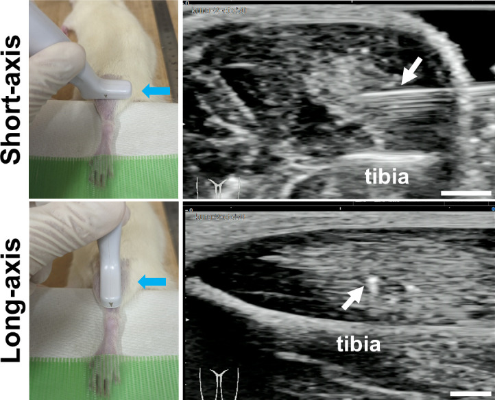
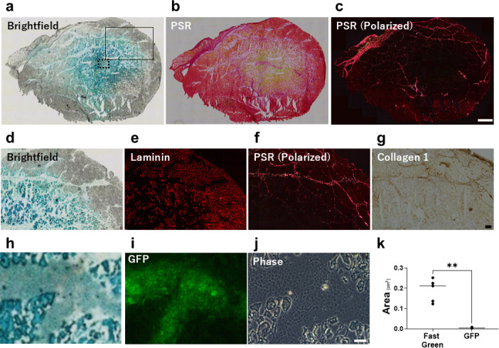
A cardiotoxin (CTX)-induced muscle injury model was used. Briefly, CTX was injected into tibialis anterior muscle in rats under ultrasound observation. First, the diagnostic accuracy of abnormal muscle lesions on ultrasound was examined by comparing ultrasound findings and histology. Next, Fast Green solution and green fluorescent protein (GFP)-labelled cells were simultaneously injected into the abnormal muscle lesions under ultrasound guidance, and their distribution was evaluated.
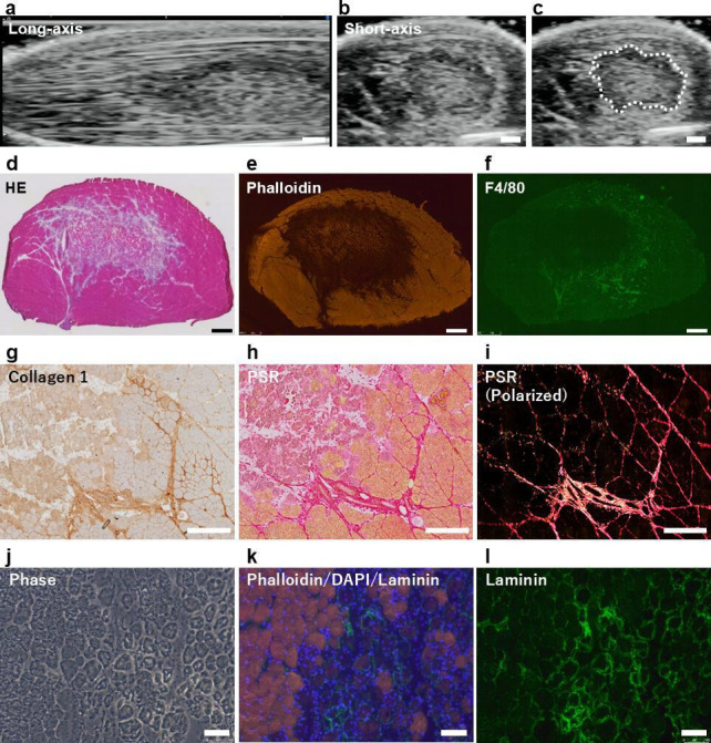
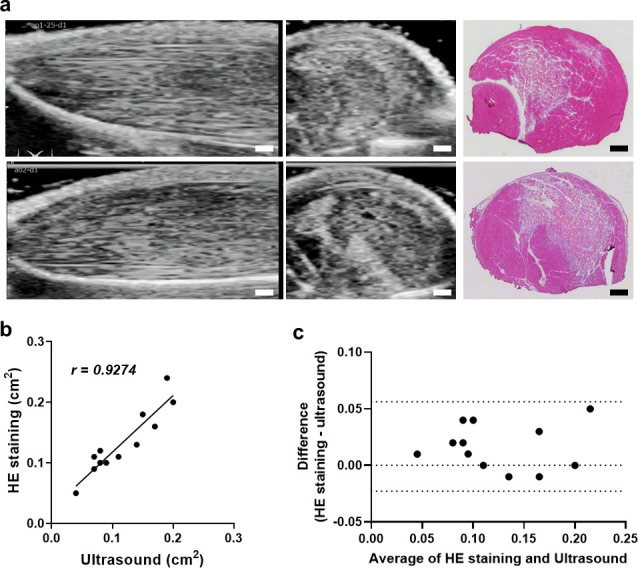
Evaluation of short-axis ultrasound images and cross-sectional histological staining showed a strong correlation (r = 0.927; p < 0.001) between the maximum muscle damage area in ultrasound and haematoxylin and eosin (H&E) staining evaluations. Histological analysis showed that ultrasound-guided injection could successfully deliver Fast Green solution around the myofibres at the site of injury. In contrast, the distribution of injected cells was very localized compared to the area stained with Fast Green.
This experimental animal study demonstrated the potential of ultrasound to quantitatively visualize abnormalities of skeletal muscle. It also showed that ultrasound-guided injections allowed for highly accurate distribution of solution and cells in abnormal muscle tissue, but the patterns of solution and cell distribution were markedly different. Although future studies using a more clinically relevant model are necessary, these results are important findings when considering biological therapies for skeletal muscle injuries and disorders.
超声引导注射技术有望提高骨骼肌损伤和疾病的治疗效果,但相关基础知识尚缺。本研究旨在探讨超声对异常骨骼肌病变的诊断准确性,并研究在超声引导下注入异常肌肉病变中的溶液和细胞的分布模式。
采用心脏毒素(CTX)诱导的肌肉损伤模型。简要来说,在超声观察下将CTX注入大鼠胫前肌。首先,通过比较超声检查结果和组织学来检测超声对异常肌肉病变的诊断准确性。接下来,在超声引导下将固绿溶液和绿色荧光蛋白(GFP)标记的细胞同时注入异常肌肉病变中,并评估它们的分布情况。
短轴超声图像评估与横断面组织学染色显示,超声检查中最大肌肉损伤面积与苏木精和伊红(H&E)染色评估结果之间存在很强的相关性(r = 0.927;p < 0.001)。组织学分析表明,超声引导注射能够成功地将固绿溶液输送到损伤部位的肌纤维周围。相比之下,与固绿染色区域相比,注入细胞的分布非常局限。
这项实验动物研究证明了超声对骨骼肌异常进行定量可视化的潜力。研究还表明,超声引导注射能够使溶液和细胞在异常肌肉组织中实现高度精确的分布,但溶液和细胞的分布模式明显不同。尽管有必要使用更具临床相关性的模型进行进一步研究,但在考虑骨骼肌损伤和疾病的生物治疗时,这些结果是重要的发现。