Li Zhuoheng, Li Juan, He Shuli, Chen Jun, Deng Chengjun, Duan Jintao
Gastroenterology Department Kunming Children's Hospital Kunming China.
Food Sci Nutr. 2025 Jan 19;13(1):e4694. doi: 10.1002/fsn3.4694. eCollection 2025 Jan.
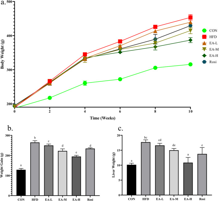
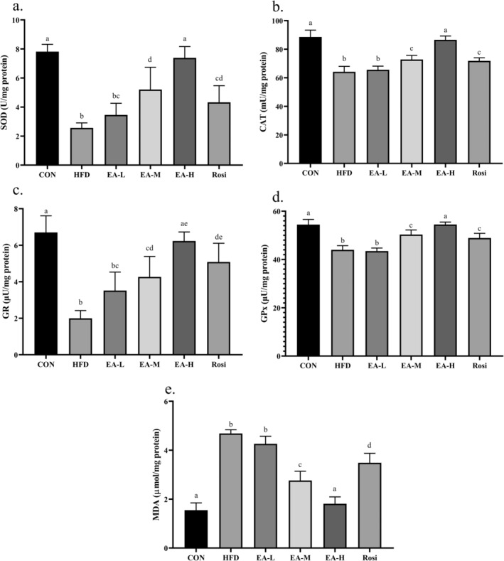
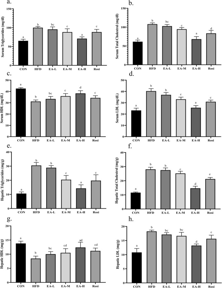
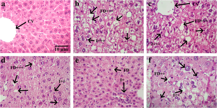
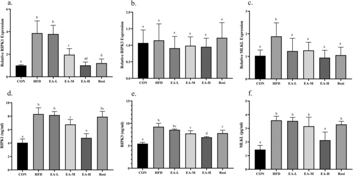
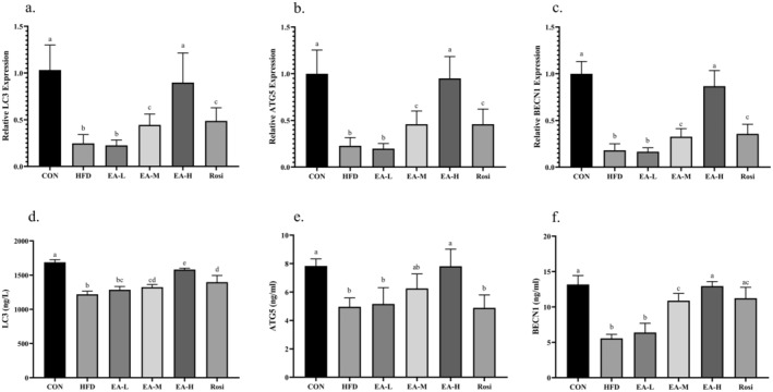
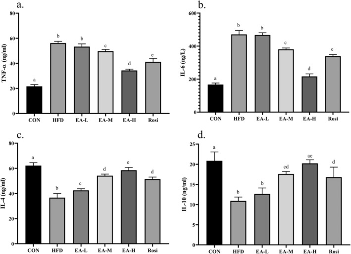
Nonalcoholic fatty liver disease (NAFLD) is considered one of the most common metabolic disorders worldwide. Although the pathoetiology of NAFLD is not fully elucidated, recent evidence suggests the involvement of stress, inflammation, and programmed death in the onset and progression of the disease. This investigation aimed to evaluate the effects of ellagic acid (EA), a known herbal antioxidant, on a high-fat diet (HFD)-induced animal model of NAFLD by evaluating the status of lipid profile, necroptosis (RIPK1, RIPK3, and MLKL), autophagy (LC3, ATG5, and BECN1), inflammation (TNF-α, IL-6, IL-4, and IL-10), and stress (SOD, CAT, GR, GPx, and MDA). In this regard, rats were randomly divided into 6 groups as follows: normal diet controls, HFD (supplemented with high caloric diet model), EA low dose (HFD and 10 mg/kg/day EA), EA middle dose (HFD and 25 mg/kg/day EA), EA high dose (HFD and 50 mg/kg/day EA), and Rosiglitazone (HFD and 10 mg/kg/day Rosi). After the treatment, the levels of markers related to necroptosis and autophagy in the liver tissue as well as the lipid profiles, inflammation, and oxidative stress status were analyzed. It was shown that the dose of EA was able to improve the weight gain and lipid profile when compared to NAFLD animals (-value < 0.001). Moreover, EA increased the level of LC3 and ATG5 while decreasing BECN 1, RIPK1, RIPK3, and MLKL compared to the HFD-induced NAFLD rats (-value < 0.05). TNF-α and IL-6 were decreased after EA administration, whereas IL-4 and IL-10 levels were increased (-value < 0.001). Furthermore, the increase in the activity of SOD, CAT, GR, and GPx along with the decrease in MDA levels indicated the suppression of oxidative stress by EA treatment compared to the NAFLD rats (-value < 0.0001). The current findings may suggest that EA improves NAFLD via modulation of necroptosis, autophagy, inflammation, and stress.
非酒精性脂肪性肝病(NAFLD)被认为是全球最常见的代谢紊乱疾病之一。尽管NAFLD的发病机制尚未完全阐明,但最近的证据表明应激、炎症和程序性死亡参与了该疾病的发生和发展。本研究旨在通过评估脂质谱、坏死性凋亡(RIPK1、RIPK3和MLKL)、自噬(LC3、ATG5和BECN1)、炎症(TNF-α、IL-6、IL-4和IL-10)以及应激(SOD、CAT、GR、GPx和MDA)的状态,来评估已知的草药抗氧化剂鞣花酸(EA)对高脂饮食(HFD)诱导的NAFLD动物模型的影响。在这方面,将大鼠随机分为6组,如下:正常饮食对照组、HFD(补充高热量饮食模型)、EA低剂量组(HFD和10mg/kg/天EA)、EA中剂量组(HFD和25mg/kg/天EA)、EA高剂量组(HFD和50mg/kg/天EA)以及罗格列酮组(HFD和10mg/kg/天罗格列酮)。治疗后,分析肝组织中与坏死性凋亡和自噬相关的标志物水平以及脂质谱、炎症和氧化应激状态。结果表明,与NAFLD动物相比,EA剂量能够改善体重增加和脂质谱(P值<0.001)。此外,与HFD诱导的NAFLD大鼠相比,EA增加了LC3和ATG5的水平,同时降低了BECN 1、RIPK1、RIPK3和MLKL的水平(P值<0.05)。EA给药后TNF-α和IL-6降低,而IL-4和IL-10水平升高(P值<0.001)。此外,与NAFLD大鼠相比,SOD、CAT、GR和GPx活性的增加以及MDA水平的降低表明EA治疗抑制了氧化应激(P值<0.0001)。目前的研究结果可能表明,EA通过调节坏死性凋亡、自噬、炎症和应激来改善NAFLD。