Zhuravlev Maxim, Kiselev Anton, Orlova Anna, Egorov Evgeniy, Drapkina Oxana, Simonyan Margarita, Drozhdeva Evgenia, Penzel Thomas, Runnova Anastasiya
Institute of Physics, Saratov State University, Astrahanskaia, 83, Saratov 410012, Russia.
National Medical Research Center for Therapy and Preventive Medicine, Petroverigsky per., 10, Moscow 101000, Russia.
Clocks Sleep. 2024 Dec 31;7(1):1. doi: 10.3390/clockssleep7010001.
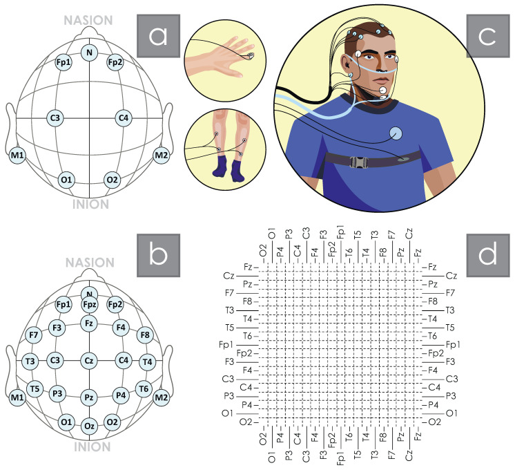
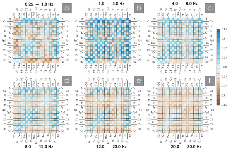
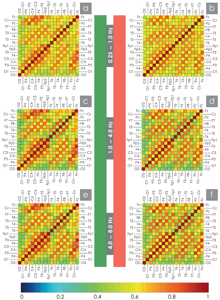
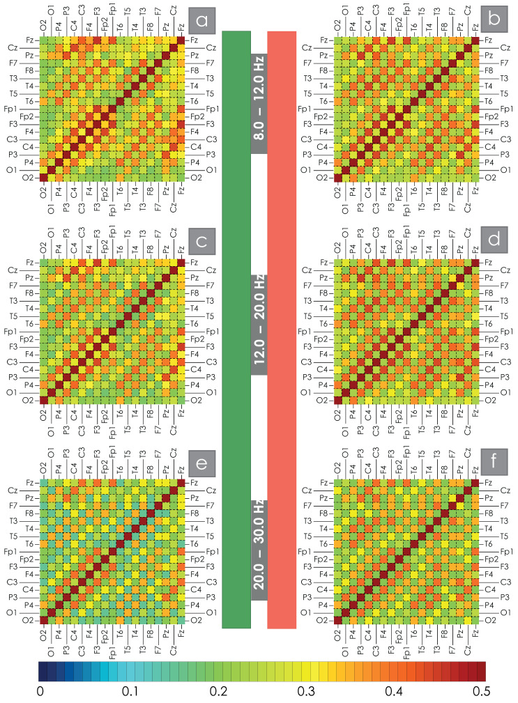
This study involved 72 volunteers divided into two groups according to the apnea-hypopnea index (AHI): AHI>15 episodes per hour (ep/h) (main group, n=39, including 28 men, median AHI 44.15, median age 47), 0≤AHI≤15ep/h (control group, n=33, including 12 men, median AHI 2, median age 28). Each participant underwent polysomnography with a recording of 19 EEG channels. Based on wavelet bicoherence (WB), the magnitude of connectivity between all pairs of EEG channels in six bands was estimated: Df1 0.25;1, Df2 1;4, Df3 4;8, Df4 8;12, Df5 12;20, Df6 20;30 Hz. In all six bands considered, we noted a significant decrease in symmetrical interhemispheric connections in OSA patients. Also, in the main group for slow oscillatory activity Df1 and Df2, we observe a decrease in connection values in the EEG channels associated with the central interhemispheric sulcus. In addition, patients with AHI>15 show an increase in intrahemispheric connectivity, in particular, forming a left hemisphere high-degree synchronization node (connections PzT3, PzF3, PzFp1) in the Df2 band. When considering high-frequency EEG oscillations, connectivity in OSA patients again shows a significant increase within the cerebral hemispheres. The revealed differences in functional connectivity in patients with different levels of AHI are quite stable, remaining when averaging the full nocturnal EEG recording, including both the entire sleep duration and night awakenings. The increase in the number of hypoxia episodes correlates with the violation of the symmetry of interhemispheric functional connections. Maximum absolute values of correlation between the apnea-hypopnea index, AHI, and the WB synchronization strength are observed for the Df2 band in symmetrical EEG channels C3C4 (-0.81) and P3P4 (-0.77). The conducted studies demonstrate the possibility of developing diagnostic systems for obstructive sleep apnea syndrome without using signals from the cardiovascular system and respiratory activity.
本研究纳入了72名志愿者,根据呼吸暂停低通气指数(AHI)分为两组:AHI>15次/小时(ep/h)(主要组,n = 39,包括28名男性,AHI中位数44.15,年龄中位数47岁),0≤AHI≤15 ep/h(对照组,n = 33,包括12名男性,AHI中位数2,年龄中位数28岁)。每位参与者均接受了包含19个脑电图通道记录的多导睡眠图检查。基于小波双相干性(WB),估计了六个频段中所有脑电图通道对之间的连接强度:Df1 0.25;1、Df2 1;4、Df3 4;8、Df4 8;12、Df5 12;20、Df6 20;30赫兹。在所有考虑的六个频段中,我们注意到阻塞性睡眠呼吸暂停(OSA)患者的对称半球间连接显著减少。此外,在主要组中,对于慢振荡活动Df1和Df2,我们观察到与中央半球间沟相关的脑电图通道中的连接值降低。另外,AHI>15的患者半球内连接增加,特别是在Df2频段形成了一个左半球高度同步节点(连接PzT3、PzF3、PzFp1)。当考虑高频脑电图振荡时,OSA患者的连接性在大脑半球内再次显著增加。在不同AHI水平的患者中揭示的功能连接差异相当稳定,在对包括整个睡眠时间和夜间觉醒在内的完整夜间脑电图记录进行平均时仍然存在。缺氧发作次数的增加与半球间功能连接对称性的破坏相关。在对称脑电图通道C3C4(-0.81)和P3P4(-0.77)的Df2频段中观察到呼吸暂停低通气指数(AHI)与WB同步强度之间的最大绝对相关值。所进行的研究表明,在不使用来自心血管系统和呼吸活动信号的情况下开发阻塞性睡眠呼吸暂停综合征诊断系统是可能的。