Yalcin Arzu, Saygin Mustafa, Ozmen Ozlem, Aslankoc Rahime, Özturk Önder, Aslancan Hasan, Kavrik Oguzhan
Department of Physiology, Faculty of Medicine, Suleyman Demirel University, Isparta, Turkey.
Department of Pathology, Burdur Mehmet Akif Ersoy University Faculty of Veterinary Medicine, Burdur, Turkey.
Iran J Basic Med Sci. 2025;28(2):264-272. doi: 10.22038/ijbms.2024.78085.16880.
This study aimed to investigate the potential effects of different doses of essential oil (Lavender EO) administered by inhalation on sleep latency and neuromodulators regulating the sleep/wake cycle in rats with total sleep deprivation (TSD).
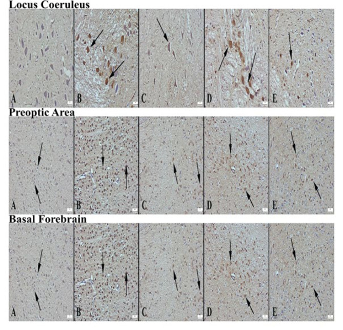
Forty-eight male Sprague-Dawley rats were divided into five groups: Control, Alprazolam (ALP, 0.25 mg/kg given intraperitoneally), L1 (Lavender EO, 0.3 ml given by inhalation), L2 (Lavender EO, 0.5 ml given by inhalation), and L3 (Lavender EO, 1 ml given by inhalation); TSD was applied to all groups. Rats in SD groups were kept on a platform surrounded by water for 18 hr for 20 days, and for the remaining time, the animals were exposed to Lavender EO for 1 hr (11:00-12:00) and then were kept in their home cage for 5 hr (12:00-17:00). Their brain and brainstem were removed for histopathological and immunohistochemical analyses (c-Fos, ChAT, GAD, and ADRB2 expression) in the locus coeruleus (LC), basal forebrain (BF), and preoptic area (PO).
The groups ranked by the severity of edema, hyperemia, and neurodegeneration in LC, BF, and PO areas were control, L3, L1, L2, and ALP. c-Fos expression significantly decreased in all brain regions in all groups except the L1 group. ChAT and GAD expressions increased dramatically in all brain regions. ADRB2 significantly increased in LC in ALP and L2 groups; in the PO area in ALP, L1, and L2 groups; and in BF in all groups.
Lavender EO treatment ameliorated c-Fos, ChAT, GAD, and ADRB2 expression, similar to the effect of ALP.
本研究旨在探讨通过吸入给予不同剂量香薰精油(薰衣草精油)对完全睡眠剥夺(TSD)大鼠的睡眠潜伏期以及调节睡眠/觉醒周期的神经调节剂的潜在影响。
48只雄性Sprague-Dawley大鼠被分为五组:对照组、阿普唑仑组(ALP,腹腔注射0.25mg/kg)、L1组(薰衣草精油,吸入0.3ml)、L2组(薰衣草精油,吸入0.5ml)和L3组(薰衣草精油,吸入1ml);所有组均施加TSD。SD组的大鼠在被水包围的平台上饲养18小时,持续20天,其余时间,动物暴露于薰衣草精油1小时(11:00 - 12:00),然后置于其饲养笼中5小时(12:00 - 17:00)。取出它们的大脑和脑干,用于蓝斑(LC)、基底前脑(BF)和视前区(PO)的组织病理学和免疫组织化学分析(c-Fos、ChAT、GAD和ADRB2表达)。
根据LC、BF和PO区域水肿、充血和神经变性的严重程度排序的组依次为对照组、L3组、L1组、L2组和ALP组。除L1组外,所有组所有脑区的c-Fos表达均显著降低。所有脑区的ChAT和GAD表达均显著增加。ALP组和L2组的LC中ADRB2显著增加;ALP组、L1组和L2组的PO区域中ADRB2显著增加;所有组的BF中ADRB2显著增加。
薰衣草精油治疗改善了c-Fos、ChAT、GAD和ADRB2的表达,类似于ALP的作用。