Turčić Marijana, Matušan Ilijaš Koviljka, Rajković Molek Koraljka, Valković Zujić Petra
Faculty of Biotehnology and Drug Research, University of Rijeka, Radmile Matejčić 2, 51000 Rijeka, Croatia.
Clinical Department of Pathology and Cytology, Clinical Hospital Centre Rijeka, Krešimirova 42, 51000 Rijeka, Croatia.
Curr Issues Mol Biol. 2025 Jan 13;47(1):47. doi: 10.3390/cimb47010047.
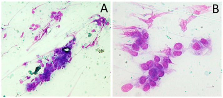
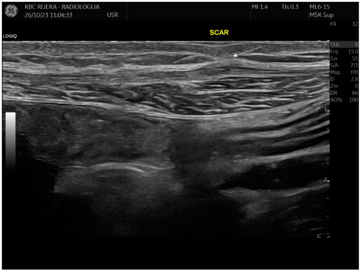
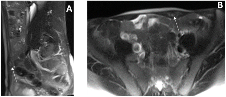
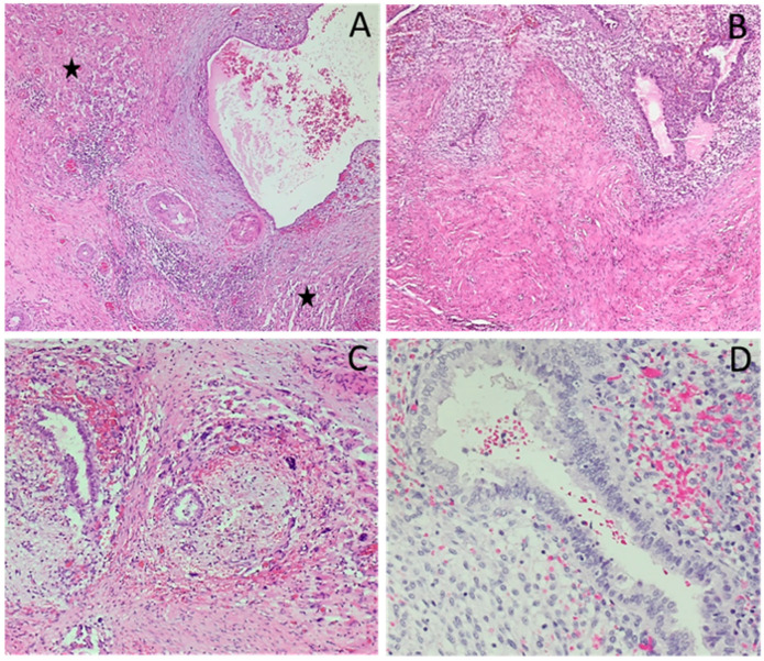
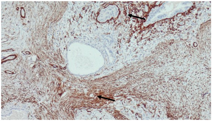
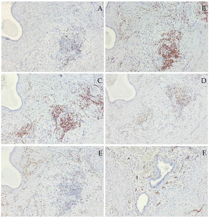
Introduction and importance: Extrapelvic endometriosis, confined exclusively to the body of the rectus abdominis muscle, is a rare form of abdominal wall endometriosis. While its etiopathology remains unclear, it is often diagnosed in healthy women who present with atypical symptoms and localization unrelated to any incision site, or in the absence of a history of endometriosis or previous surgery. Presentation of the case: Here, we describe a unique case of intramuscular endometriosis of the rectus abdominis muscle in a healthy 39-year-old Caucasian woman. The condition was located away from any prior incisional scars and presented without typical symptoms or concurrent pelvic disease, making diagnostic imaging unclear. After partial surgical resection of the endometriotic foci, the diagnosis was confirmed histologically. Progestogen-based supportive medication was initiated to prevent the need for additional surgeries and to reduce the risk of recurrence. After 6 years of follow-up and continued progestogen treatment, the patient remains symptom-free and has shown no recurrence of the disease. Clinical discussion: Endometriosis of the rectus abdominis muscle exhibits specific characteristics in terms of localization, etiopathology, symptomatology, and diagnostic imaging, suggesting that it should be considered a distinct clinical entity. Conclusions: Although rare, primary endometriosis of the rectus abdominis muscle should be included in the differential diagnosis for women of childbearing age. Early diagnosis is essential to avoid delayed recognition, tissue damage, and to minimize the risk of recurrence or malignant transformation. Given the increasing frequency of gynecologic and laparoscopic surgeries worldwide, it is crucial to establish standardized reporting protocols, follow-up timelines, and imaging assessments during specific phases of the menstrual cycle. Standardization will help raise awareness of this disease, and further our understanding of its pathogenesis, risk factors, recurrence patterns, and potential for malignant transformation-factors that are still not fully understood.
盆腔外子宫内膜异位症,仅局限于腹直肌肌腹,是腹壁子宫内膜异位症的一种罕见形式。虽然其病因病理尚不清楚,但常在健康女性中被诊断出来,这些女性表现出非典型症状且病变部位与任何手术切口部位无关,或者没有子宫内膜异位症病史或既往手术史。病例介绍:在此,我们描述了一名39岁健康白种女性腹直肌肌内子宫内膜异位症的独特病例。该病变远离任何既往手术瘢痕,且无典型症状或并发盆腔疾病,使得诊断性影像学检查结果不明确。在对子宫内膜异位病灶进行部分手术切除后,经组织学检查确诊。开始使用基于孕激素的支持性药物治疗,以避免再次手术的需要并降低复发风险。经过6年的随访和持续的孕激素治疗,患者无症状,且未出现疾病复发。临床讨论:腹直肌子宫内膜异位症在定位、病因病理、症状学和诊断性影像学方面具有特定特征,表明它应被视为一种独特的临床实体。结论:虽然罕见,但腹直肌原发性子宫内膜异位症应纳入育龄期女性的鉴别诊断中。早期诊断对于避免延迟识别、组织损伤以及将复发或恶性转化风险降至最低至关重要。鉴于全球妇科和腹腔镜手术的频率不断增加,在月经周期的特定阶段建立标准化的报告方案、随访时间表和影像学评估至关重要。标准化将有助于提高对这种疾病的认识,并加深我们对其发病机制、危险因素、复发模式和恶性转化可能性的理解,而这些因素目前仍未完全了解。