Gonzalez Mailen, Fuertes García José Manuel, Zanchetta María Belén, Abdala Rubén, Massa José María
Instituto de Investigación en Tecnología Informática Avanzada, Universidad Nacional del Centro de la Provincia de Buenos Aires, Tandil 7000, Argentina.
Consejo Nacional de Investigaciones Científicas y Técnicas, Buenos Aires 1414, Argentina.
Diagnostics (Basel). 2025 Jan 14;15(2):175. doi: 10.3390/diagnostics15020175.
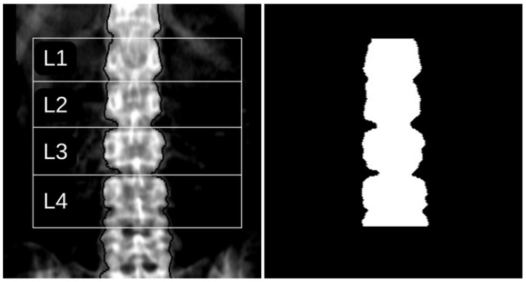
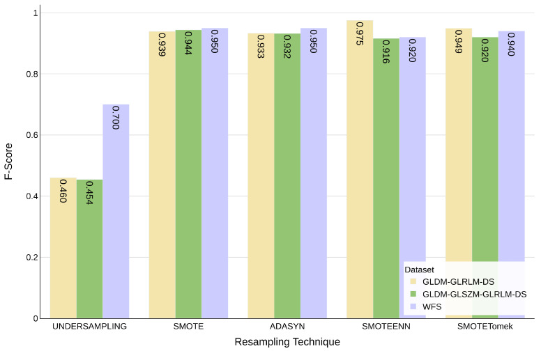
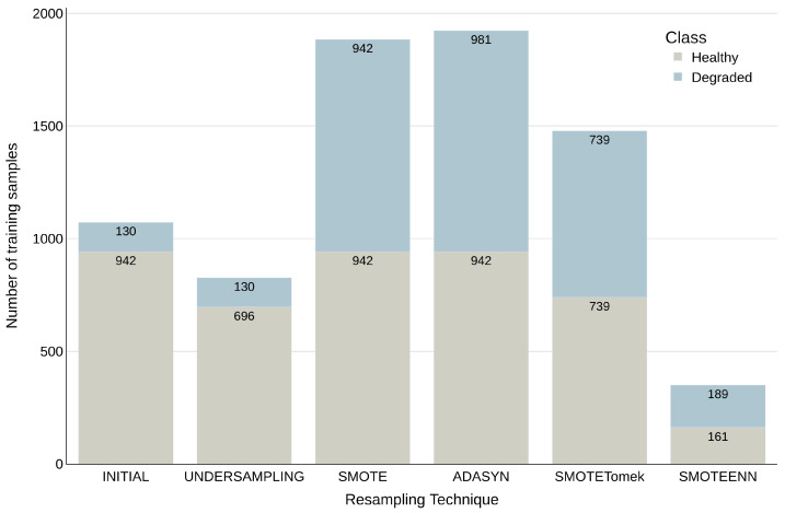
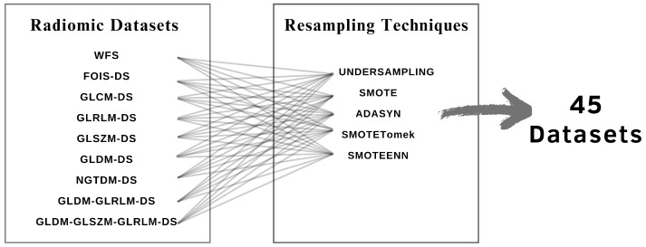
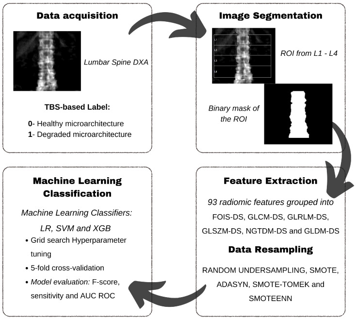
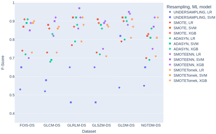
: This study presents a novel approach, based on a combination of radiomic feature extraction, data resampling techniques, and machine learning algorithms, for the detection of degraded bone structures in Dual X-ray Absorptiometry (DXA) images. This comprehensive approach, which addresses the critical aspects of the problem, distinguishes this work from previous studies, improving the performance achieved by the most similar studies. The primary aim is to provide clinicians with an accessible tool for quality bone assessment, which is currently limited. : A dataset of 1531 spine DXA images was automatically segmented and labelled based on Trabecular Bone Score (TBS) values. Radiomic features were extracted using Pyradiomics, and various resampling techniques were employed to address class imbalance. Three machine learning classifiers (Logistic Regression, Support Vector Machine (SVM), and XGBoost) were trained and evaluated using standard performance metrics. : The SVM classifier outperformed the other classifiers. The highest F-score of 97.5% was achieved using the Grey Level Dependence Matrix and Grey Level Run Length Matrix feature combination with SMOTEENN resampling, which proved to be the most effective resampling technique, while the undersampling method yielded the lowest performance. : This research demonstrates the potential of radiomic texture features, resampling techniques, and machine learning methods for classifying DXA images into healthy or degraded bone structures, which potentially leads to improved clinical diagnosis and treatment.
本研究提出了一种基于放射组学特征提取、数据重采样技术和机器学习算法相结合的新方法,用于检测双能X线吸收法(DXA)图像中的骨质退化结构。这种全面的方法解决了该问题的关键方面,使这项工作有别于以往的研究,提高了与最相似研究相比所取得的性能。主要目的是为临床医生提供一种目前有限的、易于使用的优质骨评估工具。
基于小梁骨评分(TBS)值,对1531张脊柱DXA图像的数据集进行了自动分割和标记。使用Pyradiomics提取放射组学特征,并采用各种重采样技术来解决类别不平衡问题。使用标准性能指标对三种机器学习分类器(逻辑回归、支持向量机(SVM)和XGBoost)进行了训练和评估。
SVM分类器的表现优于其他分类器。使用灰度共生矩阵和灰度游程长度矩阵特征组合以及SMOTEENN重采样(这被证明是最有效的重采样技术)时,获得了97.5%的最高F分数,而欠采样方法的性能最低。
本研究证明了放射组学纹理特征、重采样技术和机器学习方法在将DXA图像分类为健康或退化骨质结构方面的潜力,这可能会改善临床诊断和治疗。