Pushchina Evgeniya V, Pimenova Evgeniya A, Kapustyanov Ilya A, Bykova Mariya E
A.V. Zhirmunsky National Scientific Center of Marine Biology, Far Eastern Branch, Russian Academy of Sciences, 690041 Vladivostok, Russia.
Int J Mol Sci. 2025 Jan 14;26(2):644. doi: 10.3390/ijms26020644.
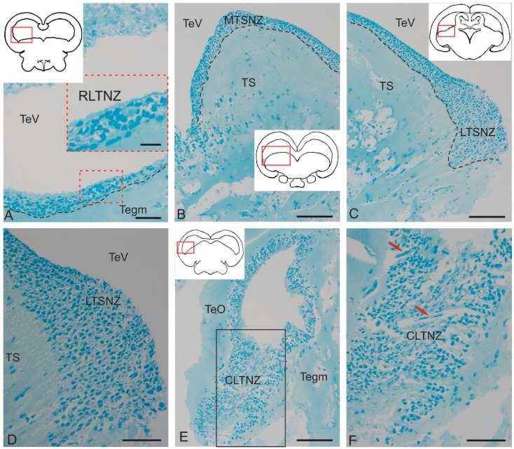
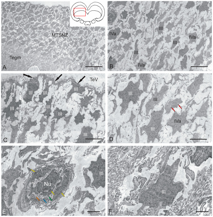
The ultrastructural organization of the nuclei of the tegmental region in juvenile chum salmon () was examined using transmission electron microscopy (TEM). The dorsal tegmental nuclei (DTN), the nucleus of (NFLM), and the nucleus of the oculomotor nerve (NIII) were studied. The ultrastructural examination provided detailed ultrastructural characteristics of neurons forming the tegmental nuclei and showed neuro-glial relationships in them. Neurons of three size types with a high metabolic rate, characterized by the presence of numerous mitochondria, polyribosomes, Golgi apparatus, and cytoplasmic inclusions (vacuoles, lipid droplets, and dense bodies), were distinguished. It was found that large interneurons of the NFLM formed contacts with protoplasmic astrocytes. Excitatory synaptic structures were identified in the tegmentum and their detailed characteristic are provided for the first time. Microglia-like cells were found in the NIII. The ultrastructural characteristics of neurogenic zones of the tegmentum of juvenile chum salmon were also determined for the first time. In the neurogenic zones of the tegmentum, adult-type neural stem progenitor cells (aNSPCs) corresponding to cells of types III and IVa Danio rerio. In the neurogenic zones of the tegmentum, neuroepithelial-like cells (NECs) corresponding to cells previously described from the zebrafish cerebellum were found and characterized. In the tegmentum of juvenile chum salmon, patterns of paracrine neurosecretion were observed and their ultrastructural characteristics were recorded. Patterns of apoptosis in large neurons of the tegmentum were examined by TEM. Using immunohistochemical (IHC) labeling of the brain lipid-binding protein (BLBP) and aromatase B (AroB), patterns of their expression in the tegmentum of intact animals and in the post-traumatic period after acute injury to the were characterized. The response to brainstem injury in chum salmon was found to activate multiple signaling pathways, which significantly increases the BLBP and AroB expression in various regions of the tegmentum and . However, post-traumatic patterns of BLBP and AroB localizations are not the same. In addition to a general increase in BLBP expression in the tegmental parenchyma, BLBP overexpression was observed in the rostro-lateral tegmental neurogenic zone (RLTNZ), while AroB expression in the RLTNZ was completely absent. Another difference was the peripheral overexpression of AroB and the formation of dense reactive clusters in the ventro-medial zone of the tegmentum. Thus, in the post-traumatic period, various pathways were activated whose components were putative candidates for inducers of the "astrocyte-like" response in the juvenile chum salmon brain that are similar to those present in the mammalian brain. In this case, BLBP acted as a factor enhancing the differentiation of both radial glia and neurons. Estradiol from AroB+ astrocytes exerted paracrine neuroprotective effects through the potential inhibition of inflammatory processes. These results indicate a new role for neuronal aromatization as a mechanism preventing the development of neuroinflammation. Moreover, our findings support the hypothesis that BLBP is a factor enhancing neuronal and glial differentiation in the post-traumatic period in the chum salmon brain.
利用透射电子显微镜(TEM)对幼年大麻哈鱼()被盖区细胞核的超微结构组织进行了检查。研究了背侧被盖核(DTN)、外侧丘系核(NFLM)和动眼神经核(NIII)。超微结构检查提供了构成被盖核的神经元的详细超微结构特征,并显示了其中的神经胶质关系。区分出了三种具有高代谢率的大小类型的神经元,其特征是存在大量线粒体、多核糖体、高尔基体和细胞质内含物(液泡、脂滴和致密体)。发现NFLM的大型中间神经元与原浆性星形胶质细胞形成了接触。首次在被盖中鉴定出兴奋性突触结构并提供了其详细特征。在NIII中发现了小胶质样细胞。还首次确定了幼年大麻哈鱼被盖区神经发生区的超微结构特征。在被盖区的神经发生区,发现了与斑马鱼小脑先前描述的细胞相对应的成年型神经干细胞祖细胞(aNSPCs),其属于III型和IVa型。在被盖区的神经发生区,发现并鉴定了与先前从斑马鱼小脑中描述的细胞相对应的神经上皮样细胞(NECs)。在幼年大麻哈鱼的被盖中,观察到了旁分泌神经分泌模式并记录了其超微结构特征。通过TEM检查了被盖中大型神经元的凋亡模式。利用脑脂质结合蛋白(BLBP)和芳香化酶B(AroB)的免疫组织化学(IHC)标记,对完整动物和急性损伤后的创伤后时期被盖中它们的表达模式进行了表征。发现大麻哈鱼对脑干损伤的反应激活了多种信号通路,这显著增加了被盖和中各个区域的BLBP和AroB表达。然而,创伤后BLBP和AroB的定位模式并不相同。除了被盖实质中BLBP表达普遍增加外,在 rostro-外侧被盖神经发生区(RLTNZ)观察到BLBP过表达,而RLTNZ中完全没有AroB表达。另一个差异是AroB在被盖腹内侧区的外周过表达以及致密反应簇的形成。因此,在创伤后时期,各种途径被激活,其成分是幼年大麻哈鱼脑中“星形胶质细胞样”反应诱导物的假定候选者,类似于哺乳动物脑中存在的那些。在这种情况下,BLBP作为促进放射状胶质细胞和神经元分化的因子发挥作用。来自AroB+星形胶质细胞的雌二醇通过潜在抑制炎症过程发挥旁分泌神经保护作用。这些结果表明神经元芳香化作为预防神经炎症发展的机制具有新的作用。此外,我们的发现支持这样的假设,即BLBP是在大麻哈鱼脑创伤后时期促进神经元和胶质细胞分化的因子。