Ruditsky Alexander, Fisher Kalin, Tighe Kayla, B'lanton Jasmine, Ma Xinrong, Jiang Kai, Byrne Kevin, Carter-Cooper Brandon, Casildo Andrea, Passaniti Antonino, Carrier France, Lapidus Rena, Richard Katharina, Kallen Michael E, Ng Vincent Y
Department of Orthopaedics, University of Maryland, Baltimore, USA.
Translational Laboratory, University of Maryland Greenebaum Comprehensive Cancer Center, Baltimore, USA.
Cureus. 2024 Dec 24;16(12):e76334. doi: 10.7759/cureus.76334. eCollection 2024 Dec.
Circulating tumor cells and clusters (CTC) from soft-tissue sarcoma (STS) that become entrapped in the lung can form micro-metastases and lead to pulmonary metastatic disease. Many patients with localized high-risk STS later develop metastases. Radiation is effective at reducing local recurrence by eradicating microscopic infiltration and satellites in the reactive zone surrounding the primary tumor. Prophylactic lung irradiation for patients with high-risk STS is a novel concept to potentially reduce the appearance of macroscopic metastases and improve survival. A proof-of-principle study was performed based on a novel approach: prophylactic lung radiation after resection of the primary tumor to address microscopic pulmonary deposits from CTC.
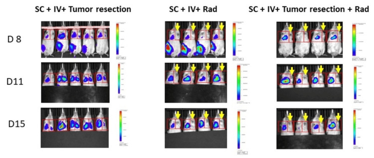
Immunocompromised mice and luciferase-expressing human fibrosarcoma (HT-1080-Luc) cell lines were used. In phase 1, HT-1080-Luc cells were injected into the tail vein to simulate CTC for the development of pulmonary metastases. Whole-lung irradiation (WLI) was then performed in the treated mice prior to the appearance of macroscopic metastases. In phase 2, a flank tumor was established to simulate a primary STS, followed by a tail-vein injection of HT-1080-Luc cells. Treatment groups included surgical removal of the primary STS and hemithoracic irradiation (HTI). Body weight and bioluminescence data were obtained and the mice were euthanized on Day 31 (phase 1) and Day 15 (phase 2) or when they reached 20% weight loss.
In phase 1, prophylactic WLI increased survival and decreased pulmonary metastases. In phase 2, prophylactic HTI (left lung) decreased pulmonary metastases compared to controls. Lung histology showed dramatically decreased growth and number of established metastases with HTI. Resection of the primary tumor did not affect the growth of metastases.
Prophylactic WLI after resection of the primary tumor to inhibit the growth of pulmonary metastases from previously entrapped CTCs may be a promising approach to improve survival for patients with localized high-risk STS.
软组织肉瘤(STS)来源的循环肿瘤细胞和细胞团(CTC)滞留在肺中可形成微转移灶并导致肺转移性疾病。许多局部高危STS患者随后会发生转移。放射治疗通过根除原发肿瘤周围反应区内的微小浸润灶和卫星灶,在降低局部复发方面有效。对高危STS患者进行预防性肺部照射是一个潜在可减少肉眼可见转移灶出现并提高生存率的新概念。基于一种新方法进行了一项原理验证研究:在切除原发肿瘤后进行预防性肺部放疗,以处理来自CTC的微小肺部沉积物。
使用免疫受损小鼠和表达荧光素酶的人纤维肉瘤(HT-1080-Luc)细胞系。在第1阶段,将HT-1080-Luc细胞注入尾静脉以模拟发生肺转移的CTC。然后在肉眼可见转移灶出现之前对治疗小鼠进行全肺照射(WLI)。在第2阶段,建立侧腹肿瘤以模拟原发性STS,随后尾静脉注射HT-1080-Luc细胞。治疗组包括手术切除原发性STS和半胸照射(HTI)。获取体重和生物发光数据,并在第31天(第1阶段)和第15天(第2阶段)或当体重减轻达到20%时对小鼠实施安乐死。
在第1阶段,预防性WLI提高了生存率并减少了肺转移。在第2阶段,与对照组相比,预防性HTI(左肺)减少了肺转移。肺组织学显示HTI使已形成转移灶的生长和数量显著减少。切除原发性肿瘤不影响转移灶的生长。
在切除原发性肿瘤后进行预防性WLI以抑制先前滞留的CTC引起的肺转移灶生长,可能是提高局部高危STS患者生存率的一种有前景的方法。