Nguyen Nhan Thanh, Le Son Hoang, Nguyen Bich-Ly Thi
Department of Oral Surgery, Faculty of Odonto-Stomatology, University of Medicine and Pharmacy at Ho Chi Minh City, Ho Chi Minh City, Viet Nam.
J Dent Sci. 2025 Jan;20(1):553-559. doi: 10.1016/j.jds.2024.04.026. Epub 2024 May 4.
BACKGROUND/PURPOSE: Autologous dentin materials are among the most promising bone substitutes for preventing osseous defects on the distal side of the lower second molar. This study aimed to investigate the effects of autologous demineralized dentin matrix on postoperative complications and wound healing after lower third molar surgery.
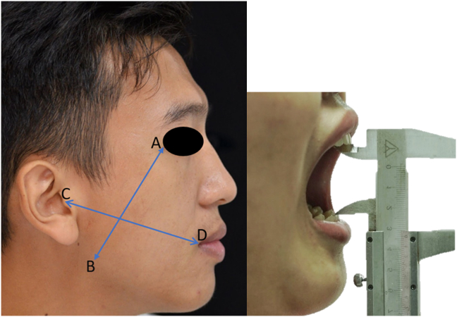
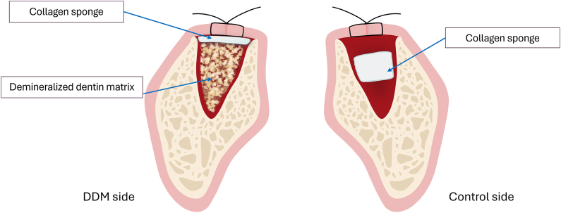
Thirteen patients with bilateral symmetrical lower third molars participated in this split-mouth randomized clinical trial. After removal surgery, one socket of the lower third molar was grafted with dentin material (demineralized dentin matrix side), and a piece of collagen sponge was used for the tooth socket of the remaining side (control side). The upper third molar on the same lateral side was extracted immediately before lower third molar surgery and used to create a demineralized dentin matrix according to the manufacturer's protocol (KometaBio). After lower third molar surgery, pain, swelling, trismus, and Inflammatory Proliferative Remodeling Scale scores were used to evaluate postoperative complications and wound healing.
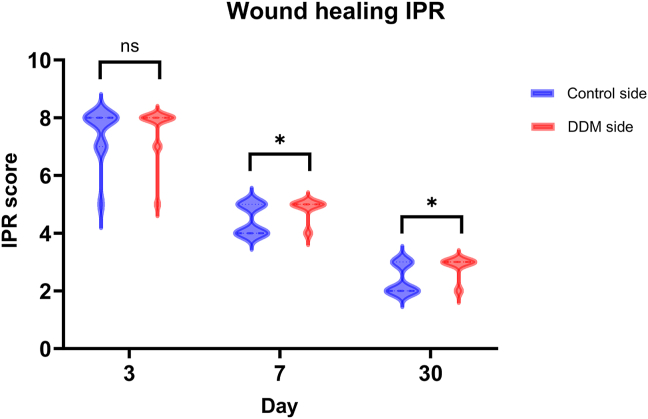
Pain, swelling, and trismus of the demineralized dentin matrix and control sides were not significantly different at any assessment time ( > 0.05). The wound-healing scores of the demineralized dentin matrix side were better than those of the control side; however, the differences were only significant at 7th and 30th days ( < 0.05).
Grafting autologous demineralized dentin matrix into the tooth socket did not increase the postoperative complications after lower third molar surgery. However, wound healing on the graft side was comparatively better than that on the control side.
NCT06073639, date 10 October, 2023. This is a retrospectively registered trial.
背景/目的:自体牙本质材料是预防下颌第二磨牙远中侧骨缺损最有前景的骨替代物之一。本研究旨在探讨自体脱矿牙本质基质对下颌第三磨牙手术后并发症及伤口愈合的影响。
13例双侧对称下颌第三磨牙患者参与了这项双侧对照随机临床试验。拔除手术后,一侧下颌第三磨牙牙槽窝植入牙本质材料(脱矿牙本质基质侧),另一侧牙槽窝使用一块胶原海绵(对照侧)。在拔除下颌第三磨牙手术前即刻拔除同侧上颌第三磨牙,并根据制造商方案(KometaBio)制备脱矿牙本质基质。下颌第三磨牙手术后,采用疼痛、肿胀、张口受限及炎症增殖重塑量表评分评估术后并发症及伤口愈合情况。
在任何评估时间,脱矿牙本质基质侧和对照侧的疼痛、肿胀及张口受限情况均无显著差异(P>0.05)。脱矿牙本质基质侧的伤口愈合评分优于对照侧;然而,差异仅在第7天和第30天有统计学意义(P<0.05)。
在下颌第三磨牙手术后,将自体脱矿牙本质基质植入牙槽窝不会增加术后并发症。然而,植入侧的伤口愈合相对优于对照侧。
NCT06073639,日期为2023年10月10日。这是一项回顾性注册试验。