Kondo Takeru, Kakinuma Hiroaki, Ambo Sara, Otake Koki, Sato Yumi, Egusa Hiroshi
Division of Molecular & Regenerative Prosthodontics, Tohoku University Graduate School of Dentistry, Sendai, Japan.
Department of Next-generation Dental Material Engineering, Tohoku University Graduate School of Dentistry, Sendai, Japan.
J Dent Sci. 2025 Jan;20(1):586-595. doi: 10.1016/j.jds.2024.04.019. Epub 2024 Apr 30.
BACKGROUND/PURPOSE: Dual-cure resin-cements are used for various dental restorations. However, whether the curing modes of these resin-cements influence gingival inflammation remains unclear. Hence, herein, we evaluated the effects of dual-cure resin-cement curing modes on gingival cytotoxicity and inflammatory responses.
Specimens were prepared using two dual-cure resin-cements-RelyX Unicem 2 (RU) and G-CEM ONE (GO)-by light-cure or self-cure modes. Degree of conversion (DC) and monomer elution of the resin-cements were measured using Fourier-transform infrared spectroscopy and high-performance liquid chromatography, respectively. Human gingival fibroblasts (GFs) and macrophages were cultured on resin-cements, and inflammatory cytokine levels, intracellular reactive oxygen species (ROS) generation, and mitogen-activated protein (MAP) kinase activation were evaluated.
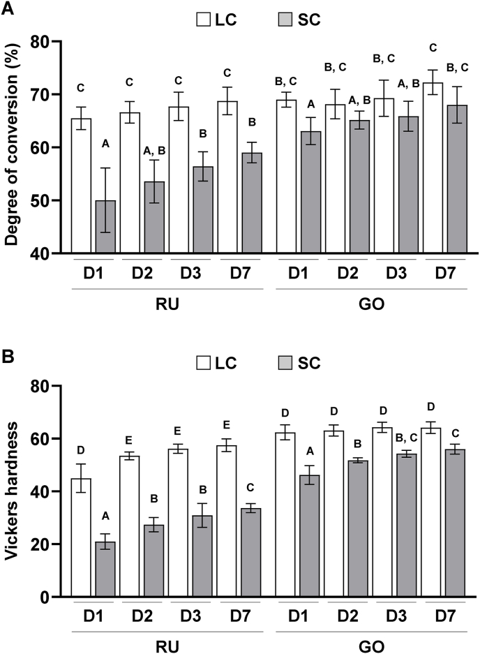
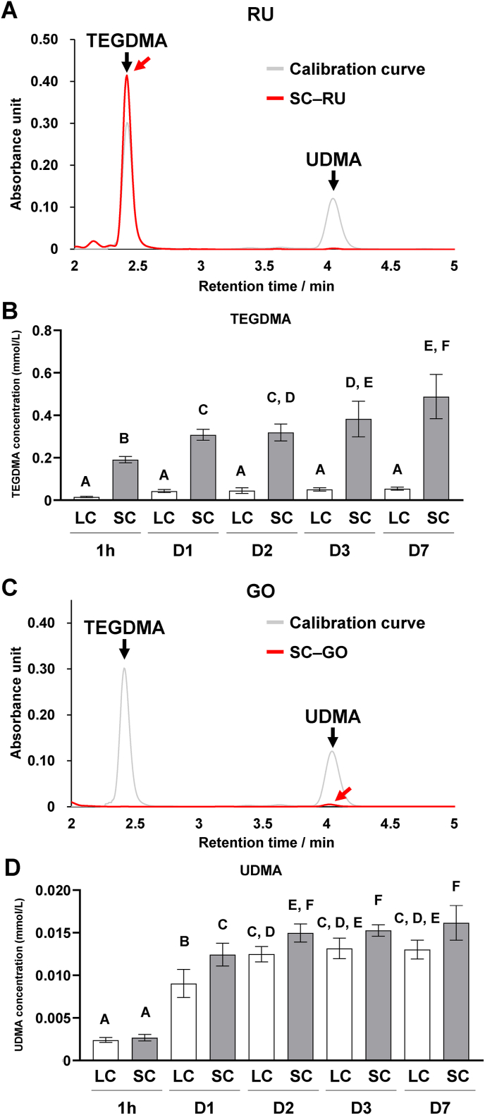
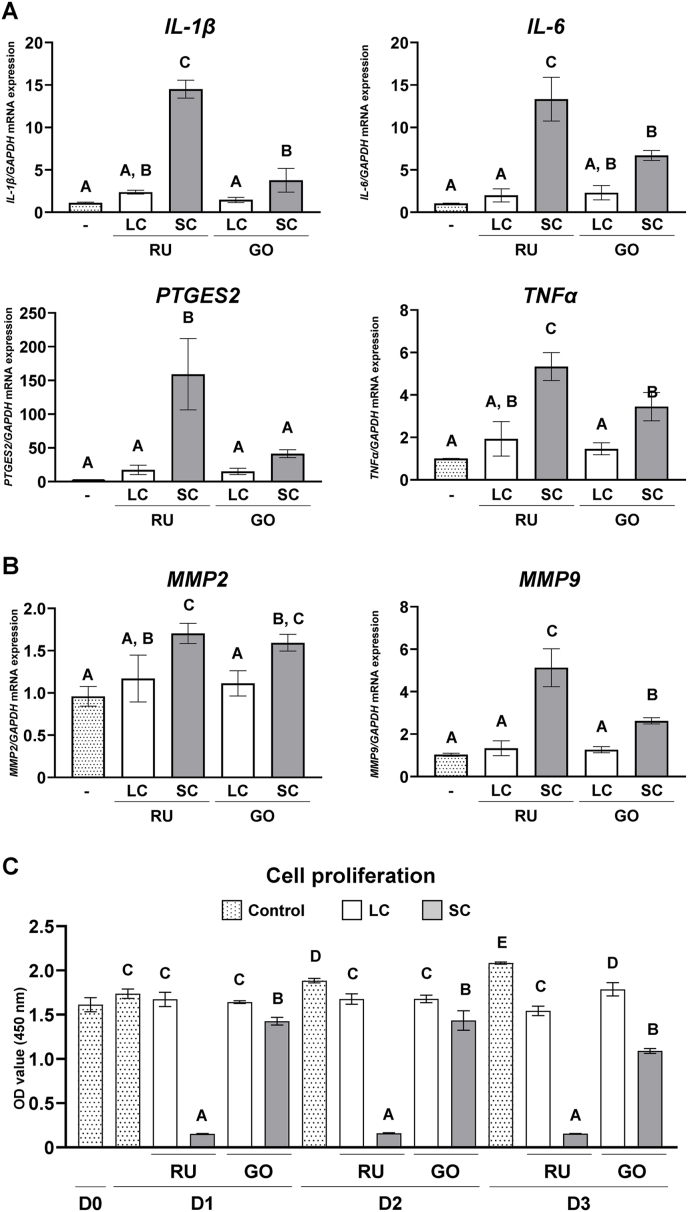
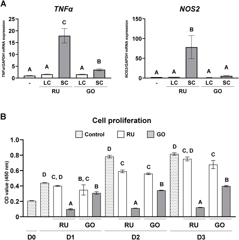
Light-cured (LC) resin-cements exhibited significantly higher DC and lower monomer elution than did self-cured (SC) resin-cements. Triethyleneglycol dimethacrylate (TEGDMA) and urethane dimethacrylate (UDMA) were substantially eluted from RU and GO, respectively. Neither LC resin-cement exhibited cytotoxicity and enhanced pro-inflammatory cytokine expression in GFs and macrophages. However, both SC resin-cements significantly decreased cell numbers and promoted cellular inflammatory responses. SC generated higher intracellular ROS levels compared to that seen with LC, and different patterns of MAP kinase activation were observed between SC-RU and SC-GO.
Compared with LC dual-cure resin-cements, SC dual-cure resin-cements show stronger cytotoxicity and elicit greater inflammatory responses in gingival cells owing to residual monomers (e.g., TEGDMA and UDMA) by activating MAP kinases in GFs and macrophages. Clinicians should ensure adequate light irradiation during prosthesis cementation and make efforts to remove the excess cement.
背景/目的:双固化树脂水门汀用于多种牙科修复。然而,这些树脂水门汀的固化模式是否会影响牙龈炎症仍不清楚。因此,在本文中,我们评估了双固化树脂水门汀固化模式对牙龈细胞毒性和炎症反应的影响。
使用两种双固化树脂水门汀——RelyX Unicem 2(RU)和G-CEM ONE(GO),通过光固化或自固化模式制备标本。分别使用傅里叶变换红外光谱和高效液相色谱法测量树脂水门汀的转化率(DC)和单体洗脱量。将人牙龈成纤维细胞(GFs)和巨噬细胞培养在树脂水门汀上,并评估炎症细胞因子水平、细胞内活性氧(ROS)生成以及丝裂原活化蛋白(MAP)激酶激活情况。
光固化(LC)树脂水门汀的DC显著高于自固化(SC)树脂水门汀,且单体洗脱量更低。三乙二醇二甲基丙烯酸酯(TEGDMA)和聚氨酯二甲基丙烯酸酯(UDMA)分别从RU和GO中大量洗脱。两种LC树脂水门汀在GFs和巨噬细胞中均未表现出细胞毒性,也未增强促炎细胞因子表达。然而,两种SC树脂水门汀均显著减少细胞数量并促进细胞炎症反应。与LC相比,SC产生更高的细胞内ROS水平,并且在SC-RU和SC-GO之间观察到不同的MAP激酶激活模式。
与LC双固化树脂水门汀相比,SC双固化树脂水门汀在牙龈细胞中表现出更强的细胞毒性,并引发更大的炎症反应,这是由于残留单体(如TEGDMA和UDMA)通过激活GFs和巨噬细胞中的MAP激酶所致。临床医生应确保在修复体粘结过程中进行充分的光照,并努力去除多余的水门汀。