Zhang Jing, Chen Xuefeng, Lv Shilin, Hao Qingqing
Hebei North University, Zhangjiakou City, Hebei Province, P.R. China.
Hebei General Hospital, Shijiazhuang City, Hebei Province, P.R. China.
PLoS One. 2025 Jan 28;20(1):e0317295. doi: 10.1371/journal.pone.0317295. eCollection 2025.
To study the effect of Dapagliflozin on ferroptosis in rabbits with chronic heart failure and to reveal its possible mechanism.
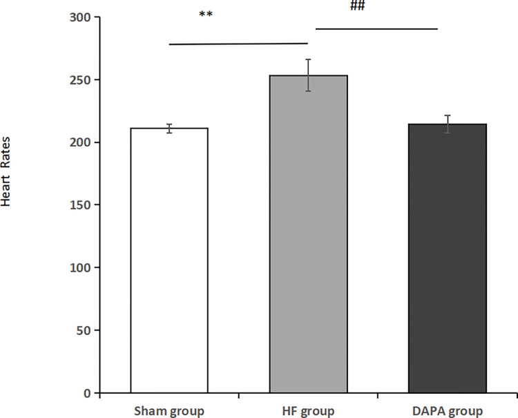
Nine healthy adult male New Zealand white rabbits were randomly divided into Sham group (only thorax opening was performed in Sham group, no ascending aorta circumferential ligation was performed), Heart failure group (HF group, ascending aorta circumferential ligation was performed in HF group to establish the animal model of heart failure), and Dapagliflozin group (DAPA group, after the rabbit chronic heart failure model was successfully made in DAPA group). Dapagliflozin was given by force-feeding method. Echocardiography was used to assess cardiac function, HE staining to evaluate pathological changes in the heart, Prussia blue staining to observe iron ions in myocardial tissue, and enzyme-linked immunosorbent assay (ELISA) to determine serum levels of the inflammatory factors interleukin-1β (IL-1β), interleukin-6 (IL-6), and tumor necrosis factor-α (TNF-α) at the end of week 12 and/or the end of week 16. The oxidative stress related indexes of malondialdehyde (MDA), superoxide dismutase (SOD) and superoxide dismutase (GSH-Px) in serum were quantitatively analyzed by colorimetry. Protein expression levels of nuclear factor E2-related factor 2(Nrf2), heme oxygenase-1(HO-1), glutathione peroxidase 4(Gpx4) were detected by Western blot.
In animals with chronic heart failure, Dapagliflozin improved cardiomyocyte hypertrophy, degeneration and necrosis. Dapagliflozin increased serum GSH-Px and SOD levels and decreased IL-1β, IL-6, TNF-α and MDA levels (P < 0.05) in a rabbit model of heart failure. Dapagliflozin also decreased cardiac iron ion levels and increased Nrf2, HO-1 and GPX4 protein expression.
Dapagliflozin can improve heart failure by inhibiting oxidative stress and ferroptosis, and its mechanism may be related to the regulation of Nrf2/HO-1/GPX4 signaling pathway.
研究达格列净对慢性心力衰竭家兔铁死亡的影响,并揭示其可能机制。
将9只健康成年雄性新西兰白兔随机分为假手术组(假手术组仅打开胸腔,不进行升主动脉环扎)、心力衰竭组(HF组,HF组进行升主动脉环扎以建立心力衰竭动物模型)和达格列净组(DAPA组,DAPA组成功制备家兔慢性心力衰竭模型后)。采用灌胃法给予达格列净。在第12周和/或第16周结束时,用超声心动图评估心功能,用苏木精-伊红(HE)染色评估心脏病理变化,用普鲁士蓝染色观察心肌组织中的铁离子,并用酶联免疫吸附测定(ELISA)法测定血清炎症因子白细胞介素-1β(IL-1β)、白细胞介素-6(IL-6)和肿瘤坏死因子-α(TNF-α)水平。采用比色法对血清中丙二醛(MDA)、超氧化物歧化酶(SOD)和谷胱甘肽过氧化物酶(GSH-Px)等氧化应激相关指标进行定量分析。通过蛋白质免疫印迹法检测核因子E2相关因子2(Nrf2)、血红素加氧酶-1(HO-1)、谷胱甘肽过氧化物酶4(Gpx4)的蛋白表达水平。
在慢性心力衰竭动物中,达格列净改善了心肌细胞肥大、变性和坏死。在心力衰竭家兔模型中,达格列净提高了血清GSH-Px和SOD水平,降低了IL-1β、IL-6、TNF-α和MDA水平(P<0.05)。达格列净还降低了心脏铁离子水平,增加了Nrf2、HO-1和GPX4蛋白表达。
达格列净可通过抑制氧化应激和铁死亡改善心力衰竭,其机制可能与调节Nrf2/HO-1/GPX4信号通路有关。