Yoshinaga Kenji, Matsushima Toma, Abe Mitsunari, Takamura Tsunehiko, Togo Hiroki, Wakasugi Noritaka, Sawamoto Nobukatsu, Murai Toshiya, Mizuno Toshiki, Matsuoka Teruyuki, Kanai Kazuaki, Hoshino Hiroshi, Sekiguchi Atsushi, Fuse Nobuo, Mugikura Shunji, Hanakawa Takashi
Department of Integrated Neuroanatomy and Neuroimaging Kyoto University Graduate School of Medicine Kyoto Japan.
Department of Advanced Neuroimaging, Integrative Brain Imaging Center National Center of Neurology and Psychiatry Kodaira Tokyo Japan.
Alzheimers Dement (Amst). 2025 Jan 29;17(1):e70048. doi: 10.1002/dad2.70048. eCollection 2025 Jan-Mar.
Brain age gap (BAG), defined as the difference between MRI-predicted 'brain age' and chronological age, can capture information underlying various neurological disorders. We investigated the pathophysiological significance of the BAG across neurodegenerative disorders.
We developed a brain age estimator using structural MRIs of healthy-aged individuals from one cohort study. Subsequently, we applied this estimator to people with Alzheimer's disease spectra (AD) and Parkinson's disease (PD) from another cohort study. We investigated brain sources responsible for BAGs among these groups.
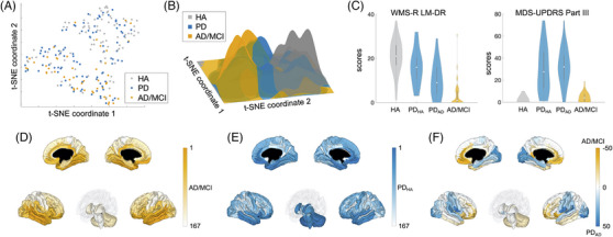
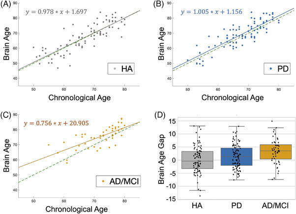
Both AD and PD exhibited a positive BAG. Brain sources showed overlapping, yet partially segregated, neuromorphological differences between these groups. Furthermore, employing with t-distributed stochastic neighbor embedding on the brain sources, we subclassified PD into two groups with and without cognitive impairment.
Our findings suggest that brain age estimation becomes a clinically relevant method for finely stratifying neurodegenerative disorders.
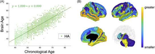
Brain age estimated from structure MRI data was greater than chronological age in patients with Alzheimer's disease/mild cognitive impairment or Parkinson's disease.Brain regions attributed to brain age estimation were located mainly in the fronto-temporo-parietal cortices but not in the motor cortex or subcortical regions.Brain sources responsible for the brain age gaps revealed roughly overlapping, yet partially segregated, neuromorphological differences between participants with Alzheimer's disease/mild cognitive impairment and Parkinson's disease.Participants with Parkinson's disease were subclassified into two groups (with and without cognitive impairment) based on brain sources responsible for the brain age gaps.
脑年龄差距(BAG)定义为磁共振成像(MRI)预测的“脑年龄”与实际年龄之间的差异,它能够捕捉各种神经系统疾病背后的信息。我们研究了BAG在神经退行性疾病中的病理生理意义。
我们利用一项队列研究中健康老年人的结构MRI数据开发了一种脑年龄估计器。随后,我们将此估计器应用于另一项队列研究中的阿尔茨海默病谱系(AD)和帕金森病(PD)患者。我们研究了这些组中导致BAG的脑源。
AD和PD均表现出正的BAG。脑源显示出这些组之间存在重叠但部分分离的神经形态学差异。此外,通过对脑源进行t分布随机邻域嵌入,我们将PD分为有认知障碍和无认知障碍两组。
我们的研究结果表明,脑年龄估计成为一种在临床上对神经退行性疾病进行精细分层的相关方法。
从结构MRI数据估计的脑年龄在阿尔茨海默病/轻度认知障碍或帕金森病患者中大于实际年龄。归因于脑年龄估计的脑区主要位于额颞顶叶皮质,而不在运动皮质或皮质下区域。导致脑年龄差距的脑源在阿尔茨海默病/轻度认知障碍参与者和帕金森病参与者之间显示出大致重叠但部分分离的神经形态学差异。基于导致脑年龄差距的脑源,帕金森病参与者被分为两组(有认知障碍和无认知障碍)。