Department of Neurology, Washington University in St. Louis, St Louis, United States.
Department of Radiology, Washington University in St. Louis, St Louis, United States.
Elife. 2023 Jan 6;12:e81869. doi: 10.7554/eLife.81869.
Estimates of 'brain-predicted age' quantify apparent brain age compared to normative trajectories of neuroimaging features. The brain age gap (BAG) between predicted and chronological age is elevated in symptomatic Alzheimer disease (AD) but has not been well explored in presymptomatic AD. Prior studies have typically modeled BAG with structural MRI, but more recently other modalities, including functional connectivity (FC) and multimodal MRI, have been explored.
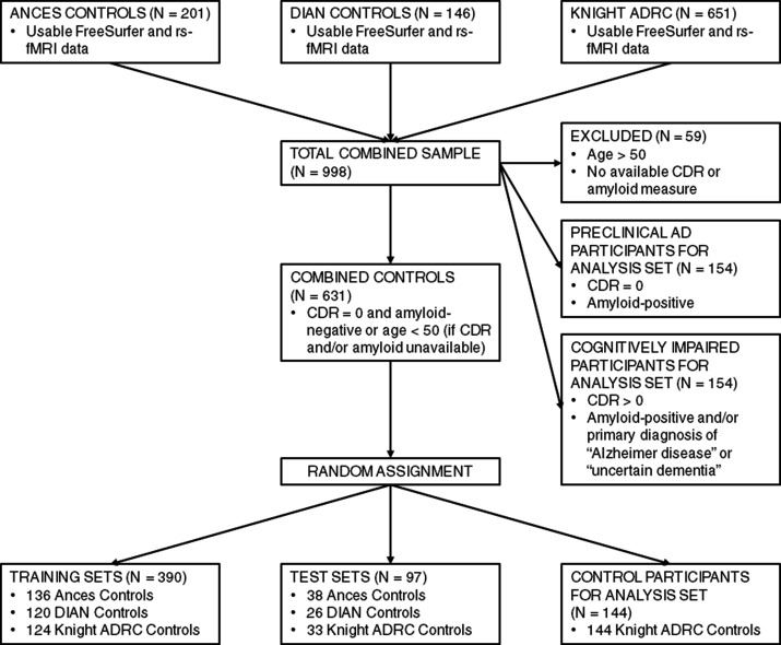
We trained three models to predict age from FC, structural (S), or multimodal MRI (S+FC) in 390 amyloid-negative cognitively normal (CN/A-) participants (18-89 years old). In independent samples of 144 CN/A-, 154 CN/A+, and 154 cognitively impaired (CI; CDR > 0) participants, we tested relationships between BAG and AD biomarkers of amyloid and tau, as well as a global cognitive composite.
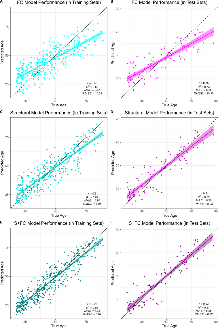
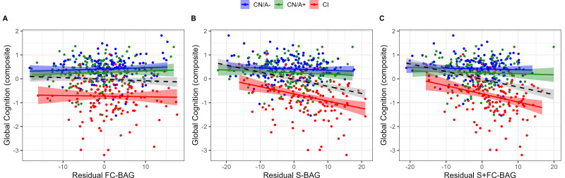
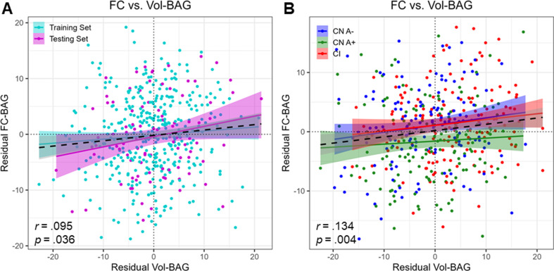
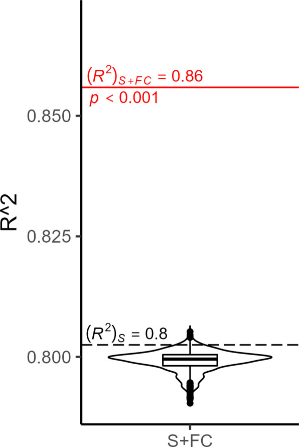
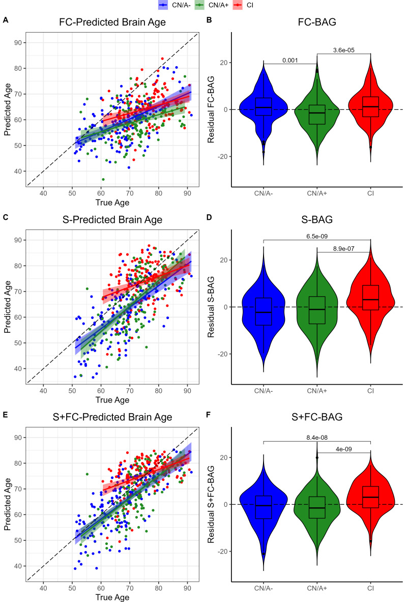
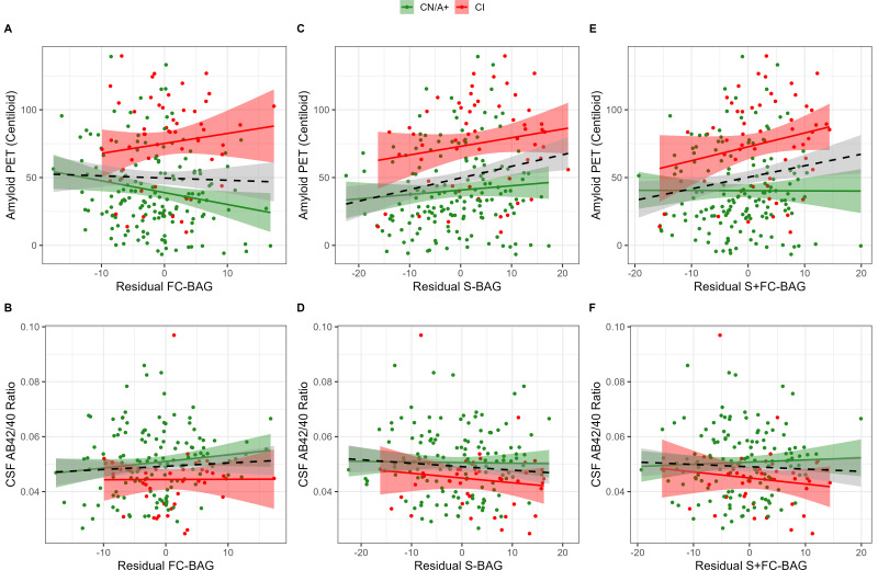
All models predicted age in the control training set, with the multimodal model outperforming the unimodal models. All three BAG estimates were significantly elevated in CI compared to controls. FC-BAG was significantly reduced in CN/A+ participants compared to CN/A-. In CI participants only, elevated S-BAG and S+FC BAG were associated with more advanced AD pathology and lower cognitive performance.
Both FC-BAG and S-BAG are elevated in CI participants. However, FC and structural MRI also capture complementary signals. Specifically, FC-BAG may capture a unique biphasic response to presymptomatic AD pathology, while S-BAG may capture pathological progression and cognitive decline in the symptomatic stage. A multimodal age-prediction model improves sensitivity to healthy age differences.
This work was supported by the National Institutes of Health (P01-AG026276, P01- AG03991, P30-AG066444, 5-R01-AG052550, 5-R01-AG057680, 1-R01-AG067505, 1S10RR022984-01A1, and U19-AG032438), the BrightFocus Foundation (A2022014F), and the Alzheimer's Association (SG-20-690363-DIAN).
“大脑预测年龄”的估计值可量化与神经影像学特征的正常轨迹相比的表观脑年龄。在有症状的阿尔茨海默病(AD)中,预测年龄与实际年龄之间的“大脑年龄差距(BAG)”升高,但在无症状 AD 中尚未得到很好的研究。先前的研究通常使用结构 MRI 来建模 BAG,但最近已经探索了其他模式,包括功能连接(FC)和多模态 MRI。
我们在 390 名淀粉样蛋白阴性认知正常(CN/A-)参与者(18-89 岁)中使用三种模型从 FC、结构(S)或多模态 MRI(S+FC)中训练预测年龄。在独立的 144 名 CN/A-、154 名 CN/A+和 154 名认知障碍(CDR>0)参与者的样本中,我们测试了 BAG 与 AD 淀粉样蛋白和 tau 的生物标志物以及全球认知综合指标之间的关系。
所有模型均在对照组的训练集中预测年龄,多模态模型的表现优于单模态模型。与对照组相比,所有三种 BAG 估计值在 CI 中均显着升高。与 CN/A-相比,CN/A+参与者的 FC-BAG 显着降低。仅在 CI 参与者中,升高的 S-BAG 和 S+FC BAG 与更高级别的 AD 病理学和更低的认知表现相关。
在 CI 参与者中,FC-BAG 和 S-BAG 均升高。然而,FC 和结构 MRI 也捕获了互补的信号。具体而言,FC-BAG 可能反映了对无症状 AD 病理学的独特双相反应,而 S-BAG 可能反映了在有症状阶段的病理性进展和认知下降。多模态年龄预测模型提高了对健康年龄差异的敏感性。
这项工作得到了美国国立卫生研究院(P01-AG026276、P01-AG03991、P30-AG066444、5-R01-AG052550、5-R01-AG057680、1-R01-AG067505、1S10RR022984-01A1 和 U19-AG032438)、BrightFocus 基金会(A2022014F)和阿尔茨海默病协会(SG-20-690363-DIAN)的支持。