Clouston Sean A P, Vaska Paul, Babalola Tesleem, Gardus John, Huang Chuan, Soriolo Nicola, Fontana Ashley, DeLorenzo Christine, Parsey Ramin, Luft Benjamin J
Program in Public Health, Stony Brook University, Stony Brook, NY, 11794, USA.
Department of Family, Population, and Preventive Medicine, Stony Brook University, Stony Brook, NY, 11794, USA.
Brain Behav Immun Health. 2025 Jan 16;44:100945. doi: 10.1016/j.bbih.2025.100945. eCollection 2025 Mar.
This study examined the regional distribution of glial activation in essential workers with neurological post-acute sequelae of coronavirus disease 2019 (COVID-19) infections (N-PASC).
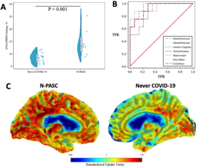
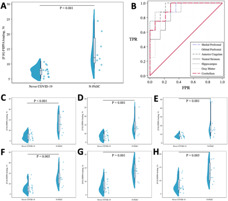
We injected ≤185 MBq of [F]-FEPPA as an intravenous bolus and positron-emission tomography over 2 h. To measure distribution volume (V) we recruited 24 essential workers (14 N-PASC, 10 Never-COVID-19 Controls, of whom 22 successfully placed arterial lines). Individuals with low binding affinity were excluded from this study, and V was adjusted for translocator protein genotype. Analyses that passed the false discovery rate are reported.
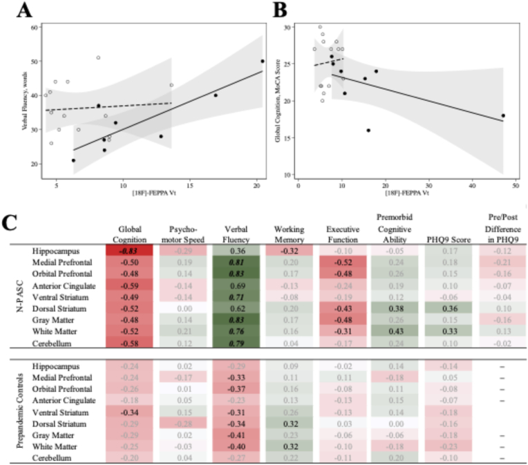
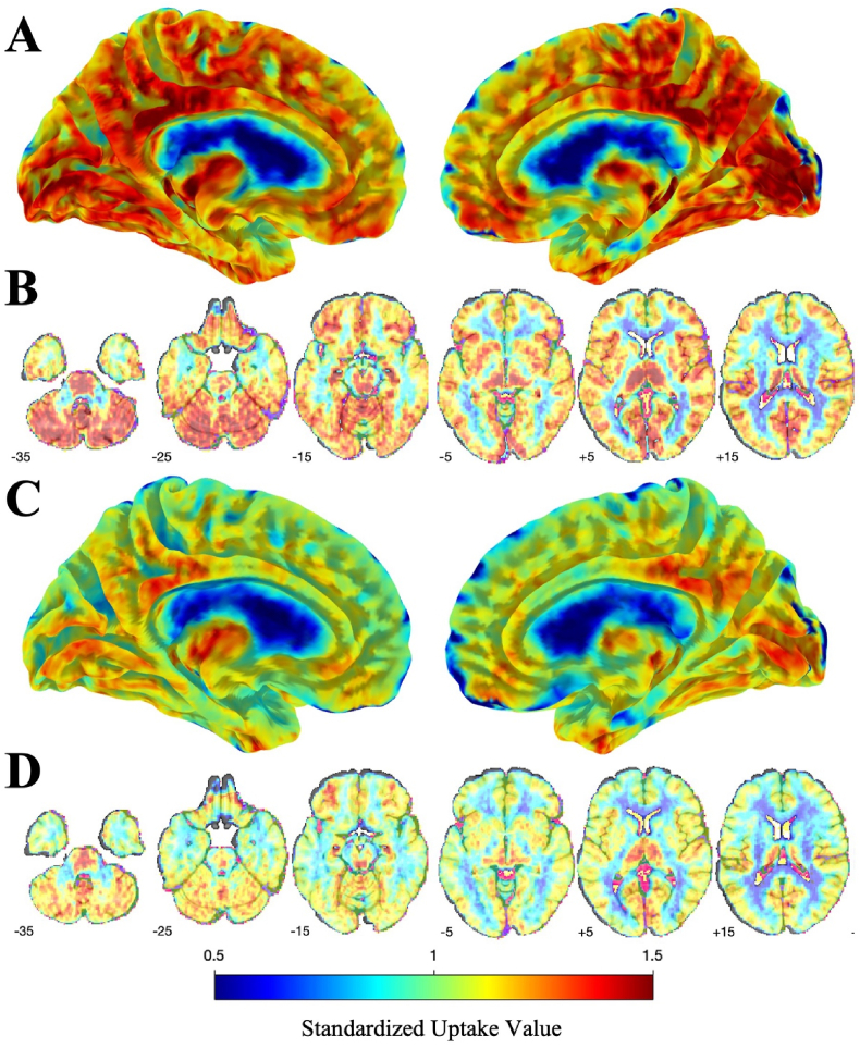
Participants at midlife survived mild to moderate COVID-19 without hospitalization but reported onset of post-acute sequelae of COVID-19 (PASC) for, on average, 22 months before undergoing neuroimaging. Hippocampal V was higher (V = 1.70, 95% C.I. = [1.30-2.21], p = 0.001) in participants with persistent brain fog after COVID-19, reflecting an increase of 10.58 mL/cm in V (area under the receiver-operating curve, AUC = 0.95 [0.85-1.00]). At a cutoff of 10.6, sensitivity/specificity/accuracy were 0.88/0.93/0.91.
The results from this study imply that neuroimmune response is a distinct and identifiable characteristic of brain fog after COVID-19. Results suggest that [F]-FEPPA could be used to support N-PASC diagnosis.
本研究调查了2019冠状病毒病(COVID-19)感染后神经后遗症(N-PASC)的一线工作者中神经胶质细胞激活的区域分布情况。
我们静脉推注≤185MBq的[F]-FEPPA,并在2小时内进行正电子发射断层扫描。为了测量分布容积(V),我们招募了24名一线工作者(14名N-PASC患者,10名从未感染过COVID-19的对照者,其中22人成功置入动脉导管)。本研究排除了低结合亲和力的个体,并根据转位蛋白基因型对V进行了调整。报告了通过错误发现率检验的分析结果。
中年参与者在未住院的情况下经历了轻度至中度COVID-19感染,但报告称在进行神经影像学检查前平均有22个月出现COVID-19急性后遗症(PASC)。COVID-19后持续存在脑雾的参与者海马体V值较高(V = 1.70,95%置信区间 = [1.30 - 2.21],p = 0.001),反映出V值增加了10.58 mL/cm(受试者工作特征曲线下面积,AUC = 0.95 [0.85 - 1.00])。截断值为10.6时,灵敏度/特异度/准确度分别为0.88/0.93/0.91。
本研究结果表明,神经免疫反应是COVID-19后脑雾的一个独特且可识别的特征。结果表明,[F]-FEPPA可用于支持N-PASC的诊断。