Meurer Steffen K, Bronneberg Gina, Penners Christian, Kauffmann Marlies, Braunschweig Till, Liedtke Christian, Huber Michael, Weiskirchen Ralf
Institute of Molecular Pathobiochemistry, Experimental Gene Therapy and Clinical Chemistry (IFMPEGKC), Medical Faculty, RWTH Aachen University, 52074, Aachen, Germany.
Institute of Biochemistry and Molecular Immunology, Medical Faculty, RWTH Aachen University, Aachen, Germany.
Cell Commun Signal. 2025 Feb 11;23(1):76. doi: 10.1186/s12964-025-02048-8.
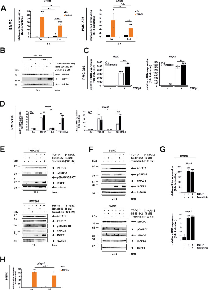
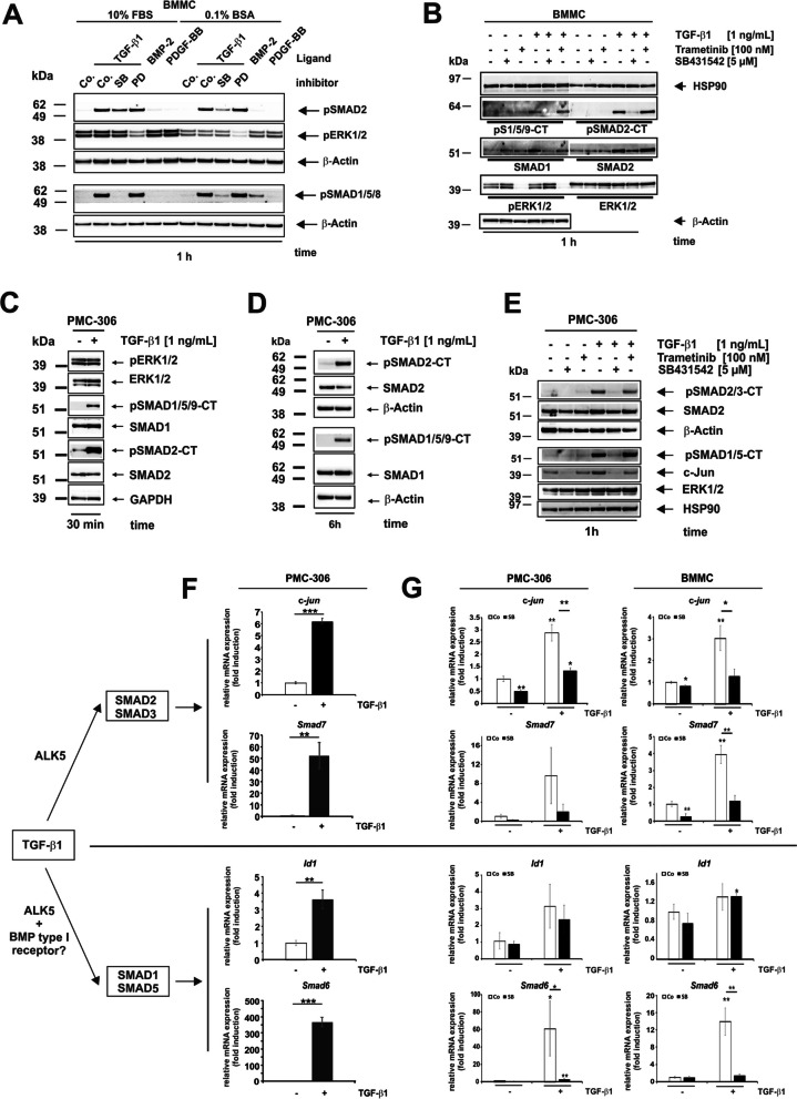
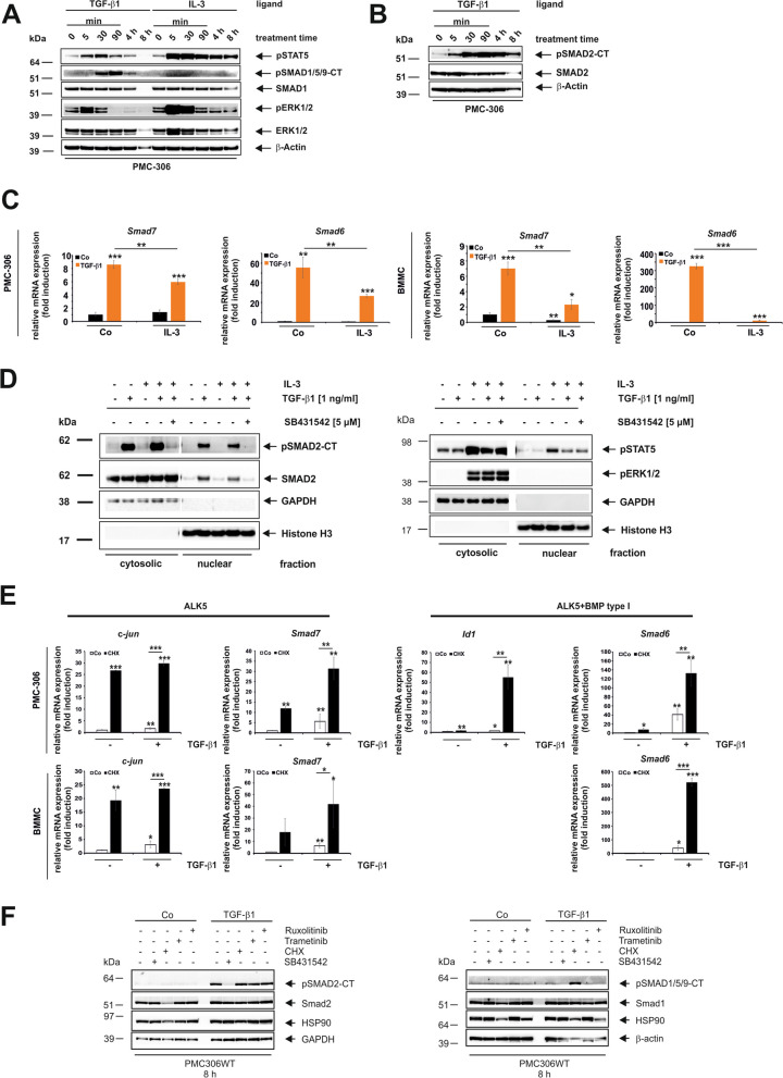
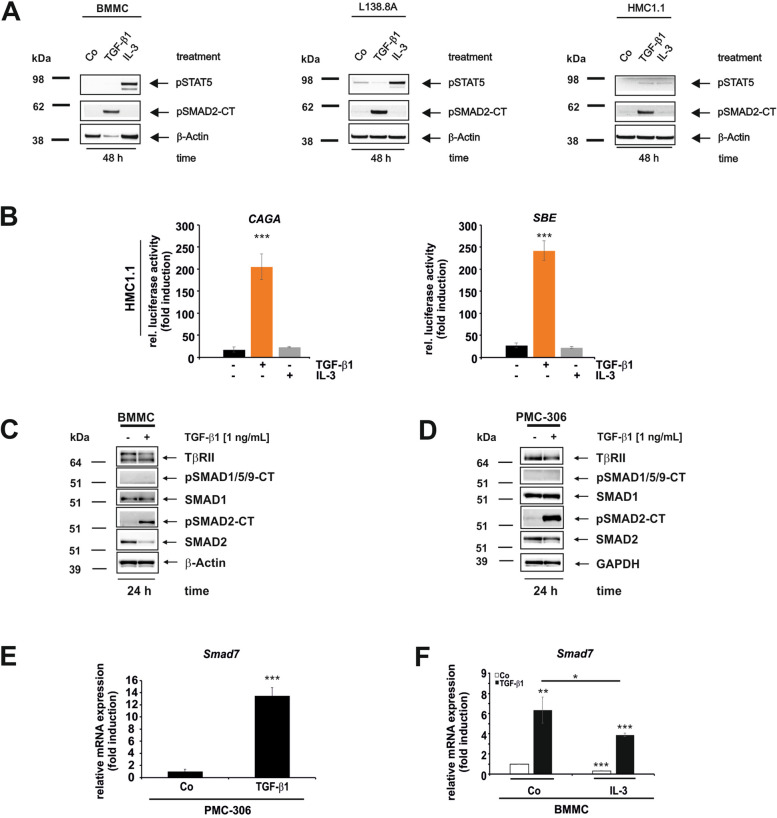
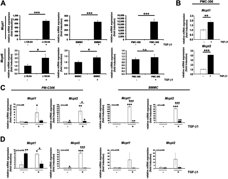
Mast cells develop from the myeloid lineage and are released from the bone marrow as immature cells, which then differentiate at the destination tissue based on cues from the local environment. In the liver, mast cells are recruited in diseased states to fibrogenic surroundings rich in TGF-β1. The aim of this study was to investigate TGF-β1 signaling in primary and permanent mast cells to identify common and unique mechanisms. The TGF-β receptor repertoire is similar among mast cells, with high expression of type I and type II receptors and very low expression of type III receptors (Betaglycan and Endoglin). Downstream, TGF-β1 activates the SMAD2/3 signaling axis and also SMAD1/5 with target genes Smad6 and Id1 in a transient manner. Initially, TGF-β1 upregulates the transcription of mucosal mast cell effectors Mcpt1 and Mcpt2 in all analyzed mast cells. This upregulation is reduced in the presence of IL-3, which promotes proliferation. Inhibition of ERK1/2 activation reduces proliferation and mitigates the negative effect of IL-3 on Mcpt1 mRNA and protein expression in the immortalized mast cell line PMC-306 but not in bone marrow-derived mast cells. Therefore, extracellular signal-regulated kinases ERK1/2 are identified as a mutual switch between IL-3-driven proliferation and TGF-β1-promoted mucosal mast cell differentiation in PMC-306. In conclusion, TGF-β1 promotes a mucosal gene signature and inhibits proliferation in mast cells, with these effects being counter-regulated by IL-3/ERK1/2.
肥大细胞起源于髓系谱系,作为未成熟细胞从骨髓中释放出来,然后根据局部环境的信号在目标组织中分化。在肝脏中,肥大细胞在疾病状态下被募集到富含转化生长因子-β1(TGF-β1)的致纤维化环境中。本研究的目的是研究原代和永生肥大细胞中的TGF-β1信号传导,以确定共同和独特的机制。肥大细胞中的TGF-β受体组成相似,I型和II型受体高表达,III型受体(β-聚糖和内皮糖蛋白)表达极低。在下游,TGF-β1以瞬时方式激活SMAD2/3信号轴以及带有靶基因Smad6和Id1的SMAD1/5。最初,TGF-β1在所有分析的肥大细胞中上调黏膜肥大细胞效应分子Mcpt1和Mcpt2的转录。在促进增殖的白细胞介素-3(IL-3)存在的情况下,这种上调作用减弱。抑制细胞外信号调节激酶ERK1/2的激活可减少增殖,并减轻IL-3对永生肥大细胞系PMC-306中Mcpt1 mRNA和蛋白表达的负面影响,但对骨髓来源的肥大细胞没有影响。因此,细胞外信号调节激酶ERK1/2被确定为PMC-306中IL-3驱动的增殖和TGF-β1促进的黏膜肥大细胞分化之间的共同开关。总之,TGF-β1促进肥大细胞中的黏膜基因特征并抑制增殖,而这些作用受到IL-3/ERK1/2的反向调节。