Lo Vercio Lucas D, Green Rebecca M, Dauter Andreas, Barretto Elizabeth C, Vidal-García Marta, Devine Jay, Marchini Marta, Robertson Samuel, Zhao Xiang, Mahika Anandita, Shakir M Bilal, Guo Sienna, Boughner Julia C, Szabo-Rogers Heather, Dean Wendy, Lander Arthur D, Marcucio Ralph S, Forkert Nils D, Hallgrímsson Benedikt
Department of Cell Biology & Anatomy, Cumming School of Medicine, University of Calgary, Calgary, AB T2N 4N1, Canada.
Alberta Children's Hospital Research Institute, University of Calgary, Calgary, AB T2N 4N1, Canada.
Development. 2025 Apr 1;152(7). doi: 10.1242/dev.204511. Epub 2025 Apr 7.
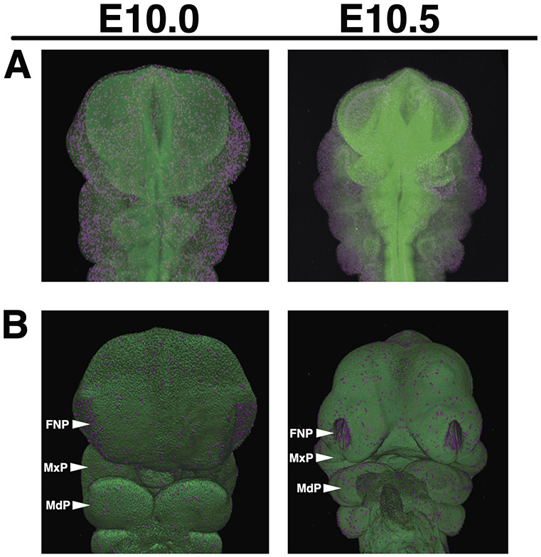
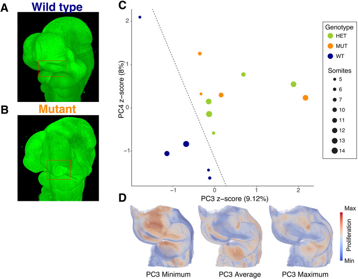
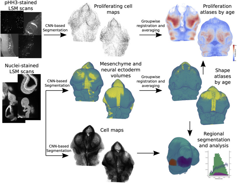
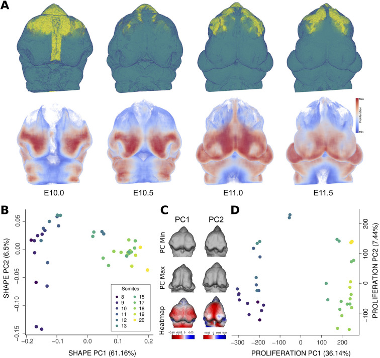
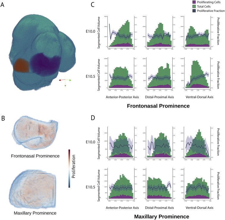
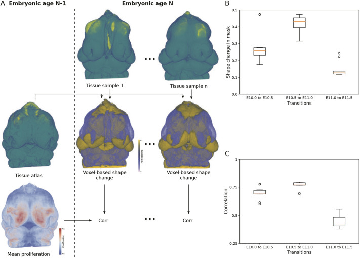
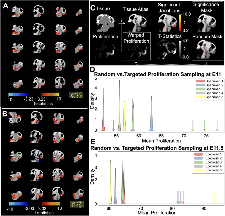
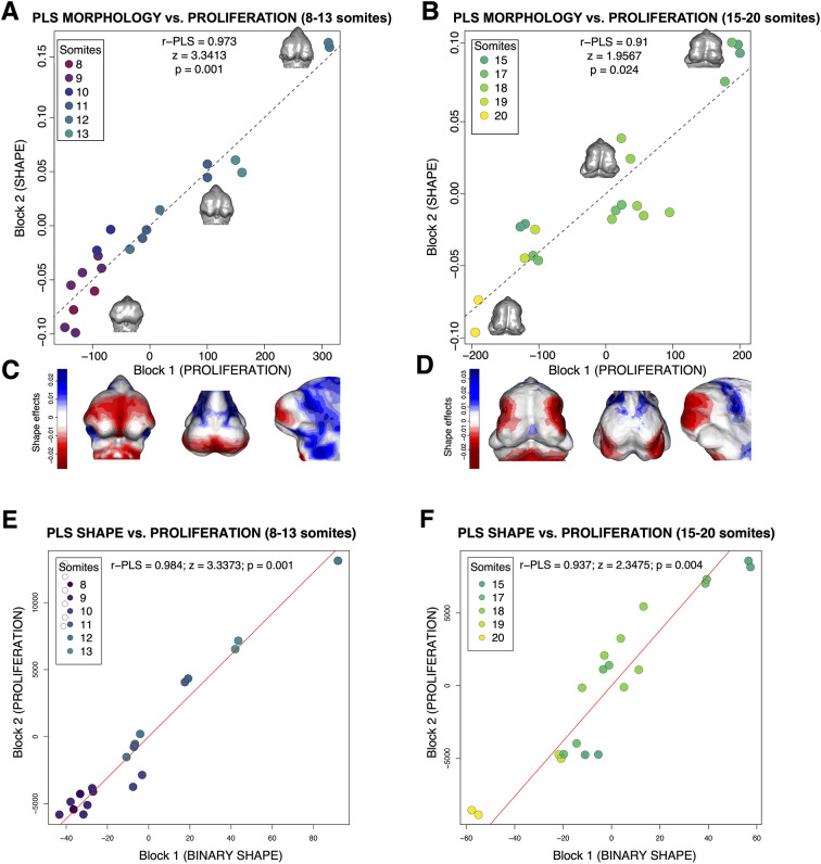
Morphogenesis requires highly coordinated, complex interactions between cellular processes: proliferation, migration and apoptosis, along with physical tissue interactions. How these cellular and tissue dynamics drive morphogenesis remains elusive. Three dimensional (3D) microscopic imaging holds great promise, and generates elegant images, but generating even moderate throughput for quantified images is challenging for many reasons. As a result, the association between morphogenesis and cellular processes in 3D developing tissues has not been fully explored. To address this gap, we have developed an imaging and image analysis pipeline to enable 3D quantification of cellular dynamics along with 3D morphology for the same individual embryo. Specifically, we focus on how 3D distribution of proliferation relates to morphogenesis during mouse facial development. Our method involves imaging with light-sheet microscopy, automated segmentation of cells and tissues using machine learning-based tools, and quantification of external morphology by geometric morphometrics. Applying this framework, we show that changes in proliferation are tightly correlated with changes in morphology over the course of facial morphogenesis. These analyses illustrate the potential of this pipeline to investigate mechanistic relationships between cellular dynamics and morphogenesis during embryonic development.
形态发生需要细胞过程之间高度协调、复杂的相互作用:增殖、迁移和凋亡,以及物理组织相互作用。这些细胞和组织动力学如何驱动形态发生仍然难以捉摸。三维(3D)显微成像前景广阔,能生成精美的图像,但由于多种原因,为定量图像生成哪怕是适度的通量都具有挑战性。因此,三维发育组织中形态发生与细胞过程之间的关联尚未得到充分探索。为了填补这一空白,我们开发了一种成像和图像分析流程,能够对同一个体胚胎的细胞动力学进行三维定量分析,并同时获取三维形态信息。具体而言,我们关注小鼠面部发育过程中增殖的三维分布与形态发生之间的关系。我们的方法包括使用光片显微镜成像、使用基于机器学习的工具对细胞和组织进行自动分割,以及通过几何形态测量学对外部形态进行定量分析。应用这个框架,我们表明在面部形态发生过程中,增殖的变化与形态的变化紧密相关。这些分析说明了这个流程在研究胚胎发育过程中细胞动力学与形态发生之间的机制关系方面的潜力。