Kan Hung-Cheng, Lin Po-Hung, Shao I-Hung, Cheng Shih-Chun, Fan Tzuo-Yau, Chang Ying-Hsu, Huang Liang-Kang, Chu Yuan-Cheng, Yu Kai-Jie, Chuang Cheng-Keng, Wu Chun-Te, Pang See-Tong, Peng Syu-Jyun
In-Service Master Program in Artificial Intelligence in Medicine, College of Medicine, Taipei Medical University, No.250, Wuxing St., Xinyi Dist., Taipei City, 110, Taiwan.
Division of Urology, Department of Surgery, Linkou Chang Gung Memorial Hospital, Taoyuan, Taiwan.
BMC Med Imaging. 2025 Feb 26;25(1):66. doi: 10.1186/s12880-025-01606-3.
This study employed a convolutional neural network (CNN) to analyze computed tomography (CT) scans with the aim of differentiating among renal tumors according to histologic sub-type.
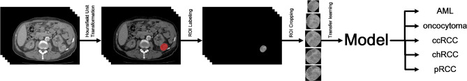
Contrast-enhanced CT images were collected from patients with renal tumors. The patient cohort was randomly split to create a training dataset (90%) and a testing dataset (10%). Following image dataset augmentation, Inception V3 and Resnet50 models were used to differentiate between renal tumors subtypes, including angiomyolipoma (AML), oncocytoma, clear cell renal cell carcinoma (ccRCC), chromophobe renal cell carcinoma (chRCC), and papillary renal cell carcinoma (pRCC). 5-fold cross validation was then used to evaluate the models in terms of classification performance.
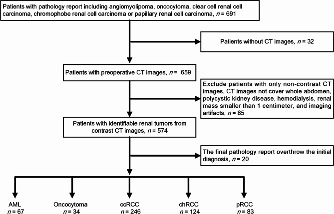
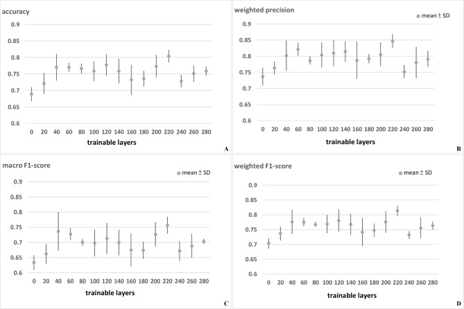
The study cohort comprised 554 patients, including those with angiomyolipoma (n = 67), oncocytoma (n = 34), clear cell renal cell carcinoma (n = 246), chromophobe renal cell carcinoma (n = 124), and papillary renal cell carcinoma (n = 83). Dataset augmentation of the training dataset included this to 4238 CT images for analysis. The accuracy of the models was as follows: Inception V3 (0.830) and Resnet 50 (0.849).
This study demonstrated the efficacy of using deep learning models for the classification of renal tumor subtypes from contrast-enhanced CT images. While the models showed promising accuracy, further development is necessary to improve their clinical applicability.
本研究采用卷积神经网络(CNN)分析计算机断层扫描(CT)图像,旨在根据组织学亚型区分肾肿瘤。
收集肾肿瘤患者的增强CT图像。将患者队列随机分为训练数据集(90%)和测试数据集(10%)。在对图像数据集进行扩充后,使用Inception V3和Resnet50模型区分肾肿瘤亚型,包括血管平滑肌脂肪瘤(AML)、嗜酸细胞瘤、透明细胞肾细胞癌(ccRCC)、嫌色细胞肾细胞癌(chRCC)和乳头状肾细胞癌(pRCC)。然后使用五折交叉验证来评估模型的分类性能。
研究队列包括554例患者,其中血管平滑肌脂肪瘤患者67例、嗜酸细胞瘤患者34例、透明细胞肾细胞癌患者246例、嫌色细胞肾细胞癌患者124例、乳头状肾细胞癌患者83例。训练数据集扩充后,分析的CT图像增加到4238张。模型的准确率如下:Inception V3为0.830,Resnet 50为0.849。
本研究证明了使用深度学习模型从增强CT图像中对肾肿瘤亚型进行分类的有效性。虽然模型显示出有前景的准确率,但仍需要进一步改进以提高其临床适用性。