Mochizuki Yuki, Joji-Nishino Asuka, Emoto Kazuo, Uematsu Akira
Department of Biological Sciences, Graduate School of Science, The University of Tokyo, Tokyo, Japan.
Human Informatics and Interaction Research Institute, National Institute for Advanced Industrial Science and Technology, Tsukuba, Japan.
Mol Brain. 2025 Mar 5;18(1):18. doi: 10.1186/s13041-025-01185-y.
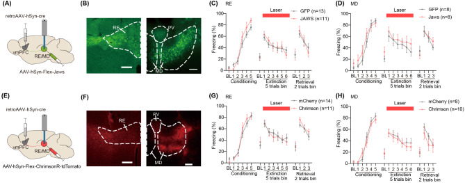
Animals adaptively regulate aversive memories in safe environments through extinction, a process central to exposure therapy for anxiety disorders. The limbic thalamus controls cognitive function in concert with interconnected cortical and limbic structures. Though medial prefrontal (mPFC) afferents to the limbic thalamus regulate aversive memory, the functional role of limbic thalamus efferents to mPFC is unclear. Here, we investigated the roles of thalamic nuclei, the reuniens (RE) and mediodorsal (MD) thalamus, projecting to the medial prefrontal cortex (mPFC) in aversive memory conditioning and extinction in male mice. Using retrograde tracing, we demonstrated that ventromedial PFC (vmPFC)- and dorsomedial PFC (dmPFC)-projecting neurons are topologically segregated within the RE and MD. Fiber photometry revealed that both RE→vmPFC and MD→vmPFC neurons respond to aversive stimuli. Notably, RE→vmPFC neurons develop shock-associated cue (CS+) response during aversive conditioning. During extinction, RE→vmPFC neurons exhibited a biphasic response to CS+, while MD→vmPFC neurons showed no cue-evoked activity. Neither optogenetic activation nor inactivation of these populations altered freezing behavior during extinction compared to controls. Collectively, these findings indicate that RE→vmPFC neurons encode aversive cue information during extinction but are dispensable for behavioral modulation. This study highlights the distinct contributions of limbic thalamus-PFC circuits to aversive memory processing.
动物在安全环境中通过消退适应性地调节厌恶记忆,这一过程是焦虑症暴露疗法的核心。边缘丘脑与相互连接的皮质和边缘结构协同控制认知功能。虽然边缘丘脑的内侧前额叶(mPFC)传入神经调节厌恶记忆,但边缘丘脑向mPFC的传出神经的功能作用尚不清楚。在这里,我们研究了投射到雄性小鼠内侧前额叶皮质(mPFC)的丘脑核团—— reunions(RE)和背内侧(MD)丘脑在厌恶记忆条件反射和消退中的作用。使用逆行追踪,我们证明了投射到腹内侧前额叶皮质(vmPFC)和背内侧前额叶皮质(dmPFC)的神经元在RE和MD内拓扑分离。纤维光度法显示,RE→vmPFC和MD→vmPFC神经元均对厌恶刺激有反应。值得注意的是,RE→vmPFC神经元在厌恶条件反射期间产生与电击相关的线索(CS+)反应。在消退过程中,RE→vmPFC神经元对CS+表现出双相反应,而MD→vmPFC神经元未表现出线索诱发的活动。与对照组相比,这些神经元群体的光遗传学激活或失活均未改变消退过程中的僵住行为。总的来说,这些发现表明,RE→vmPFC神经元在消退过程中编码厌恶线索信息,但对行为调节并非必需。这项研究突出了边缘丘脑 - PFC回路对厌恶记忆处理的不同贡献。