Talebpour Amiri Fereshteh, Omraninava Melody, Shahzamani Shadman, Khodashenas Amirali, Daryakar Arman, Nasiry Davood
Cellular and Molecular Research Center, Mazandaran University of Medical Sciences, Sari, Iran.
Hospital Administration Research Center, Sari Branch, Islamic Azad University of Medical Science, Sari, Iran.
Regen Ther. 2025 Feb 20;28:606-618. doi: 10.1016/j.reth.2025.02.002. eCollection 2025 Mar.
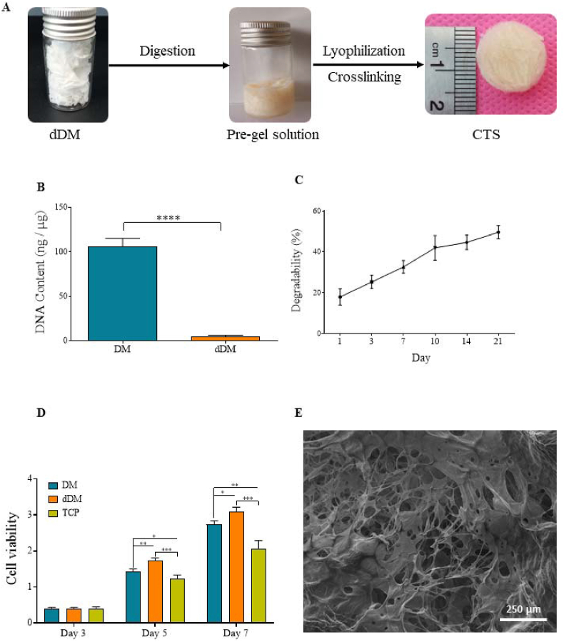
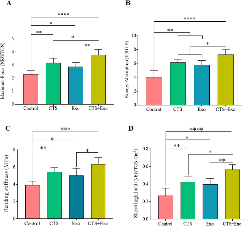
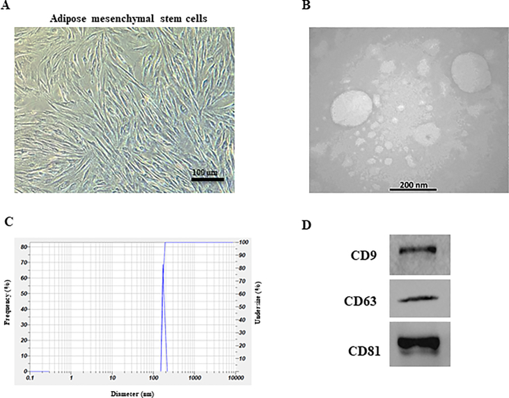
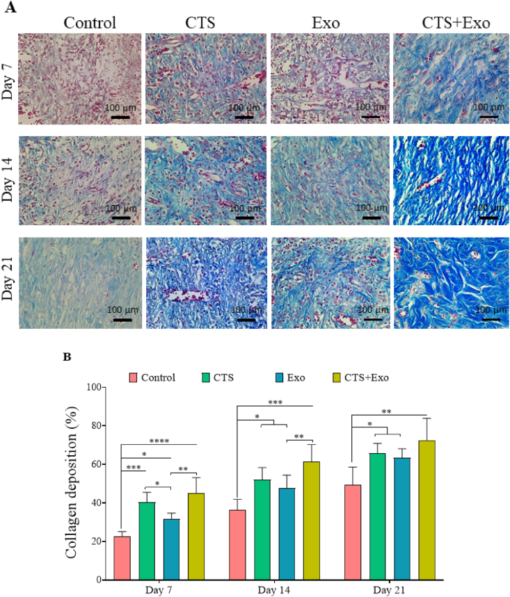
A diabetic wound is the most frequent form of chronic wound. Because diabetic wounds have multiple factors contributing to their development, the best treatments involve using a combination of approaches. Herein we assessed whether bioactive and degradable bioengineered micro-porous collagen-based three-dimensional scaffold (CTS) encapsulated with adipose mesenchymal stem cells (ASCs)-derived exosomes could accelerate the wound healing process in diabetic rats. Diabetic animals were assigned to the control group, CTS group, Exo group, and CTS+Exo group. Tissue samples were collected on days 7, 14, and 21 for evaluations including stereological, molecular, and tensiometrical assessments. The findings showed that in the treatment groups, there was a notably increase in wound closure rate, total volumes of newly formed epidermis and dermis, numerical densities of fibroblasts and blood vessels, collagen density, and biomechanical parameters than the control group, with the most noticeable changes seen in the CTS+Exo group. Additionally, there was a notably increase in the transcript of miRNA-146a, TGF-β, bFGF, and VEGF genes in the treatment groups than the control group, with the highest expression observed in the CTS+Exo group. In the CTS+Exo group, there was a much greater decrease in TNF-α and IL-1β expression, as well as in the number of neutrophils, compared to the other groups. These results validated that the combination of CTS and ASCs-derived exosomes has a greater effect on improving diabetic wound healing.
糖尿病伤口是慢性伤口最常见的形式。由于糖尿病伤口的形成有多种因素,最佳治疗方法是综合使用多种手段。在此,我们评估了包裹脂肪间充质干细胞(ASC)衍生外泌体的生物活性和可降解的生物工程微孔胶原基三维支架(CTS)是否能加速糖尿病大鼠的伤口愈合过程。将糖尿病动物分为对照组、CTS组、外泌体组和CTS+外泌体组。在第7天、14天和21天采集组织样本,进行包括体视学、分子和张力测量评估在内的评价。结果显示,与对照组相比,治疗组的伤口闭合率、新形成的表皮和真皮总体积、成纤维细胞和血管的数量密度、胶原密度以及生物力学参数均显著增加,其中CTS+外泌体组的变化最为明显。此外,与对照组相比,治疗组中miRNA-146a、TGF-β、bFGF和VEGF基因的转录本显著增加,CTS+外泌体组的表达最高。与其他组相比,CTS+外泌体组中TNF-α和IL-1β的表达以及中性粒细胞的数量有更大幅度的下降。这些结果证实,CTS和ASC衍生外泌体的组合对改善糖尿病伤口愈合有更大作用。