Hu Jiacheng, Li Yiyang, Quan Xingping, Han Yan, Chen Jinfen, Yuan Mengchen, Chen Ying, Zhou Manfei, Yu Enze, Zhou Jiahao, Wang Dawei, Wang Ruibing, Zhao Yonghua
State Key Laboratory of Quality Research in Chinese Medicine, Institute of Chinese Medical Sciences, University of Macau, Taipa, Macao SAR, People's Republic of China.
Department of Mechanical and Automation Engineering, The Chinese University of Hong Kong, Hong Kong SAR, People's Republic of China.
Chin Med. 2025 Mar 18;20(1):38. doi: 10.1186/s13020-025-01079-0.
Intravenous tissue plasminogen activator (tPA) is currently the only FDA-approved thrombolytic therapy for acute ischemic stroke (AIS), however, relative narrow therapeutic time window (within 4.5 h of AIS onset) and high risk of hemorrhagic transformation due to blood-brain barrier (BBB) disruption limit tPA therapeutic benefits for patients. In this study, we extended the time window of tPA administration (5 h after the occurrence of AIS) and investigated whether Chinese medicine classical formula Shengui Sansheng San (SSS) administration was able to alleviate BBB integrity worsening, and the mechanism was related to vasoactive intestinal peptide (VIP)/ VIP receptor 1 (VIPR1) pathway.
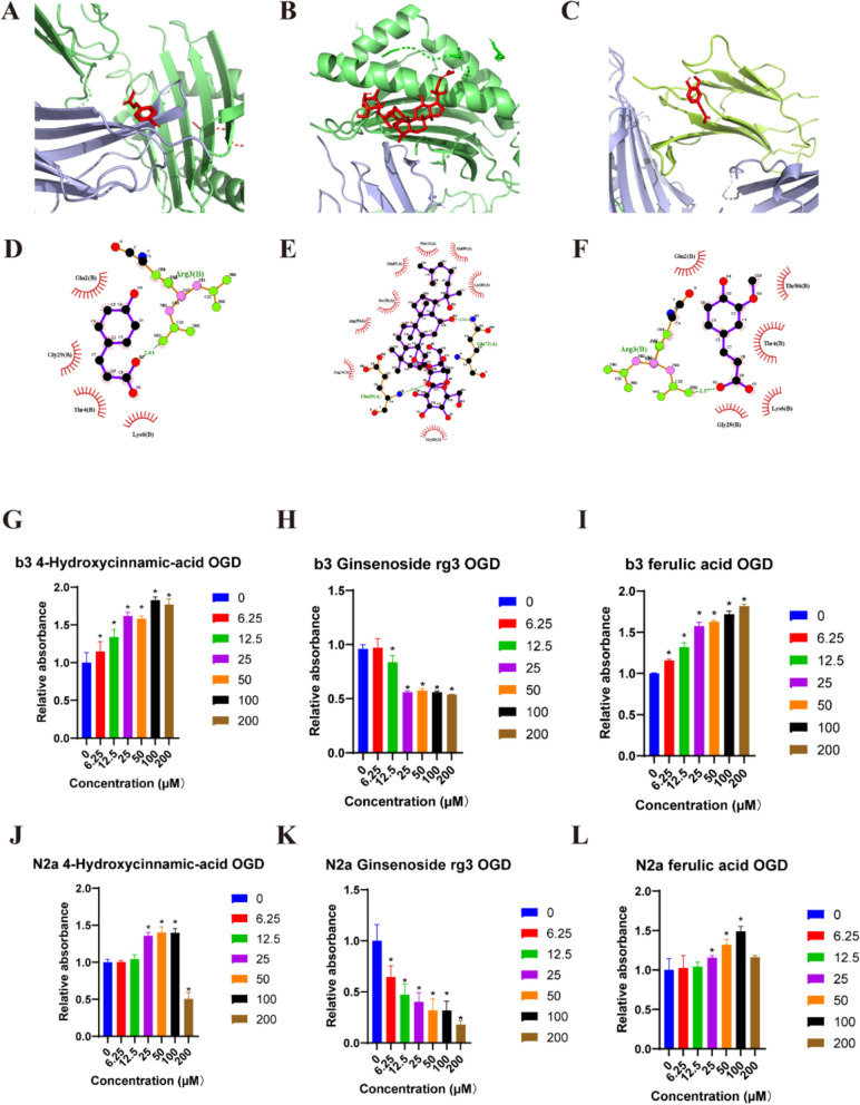
SSS was extracted using aqueous heating method and SFE-CO technology, and quality control was performed using UHPLC/MS analysis. Male C57BL/6 mice were suffered from middle cerebral artery occlusion (MCAo), followed by the removal of a silicone filament after 5 h, then, t-PA was administered via tail vein injection at once, along with SSS administration by gavage. Hemoglobin levels and Evans blue leakage were measured to assess brain hemorrhagic transformation and BBB permeability, respectively. Transmission electron microscope (TEM) was utilized to present brain microvascular endothelial cells (BMECs) tight junction morphology. TTC staining and laser speckle contrast imaging were employed for infarct volume and cerebral blood flow measurements. The modified neurological severity score (mNSS) test was conducted to evaluate neurological function. The expressions of VIP, VIPR1, ZO-1, Occludin, Lectin, GFAP, NeuN were detected by immunofluorescence staining or western blotting. In vitro, bEnd.3 and N2a cells were insulted by oxygen-glucose deprivation (OGD), and VIPR1 siRNA, and VIP shRNA transfection were respectively performed, and the molecular docking was applied to verify the SSS in-serum active compounds interacted with VIPR1. The transwell system was utilized to detect OGD-insulted BMECs permeability.
SSS treatment significantly reduced the infarct area, cerebral hemorrhage, and neurological deficits, and enhanced cerebral blood flow in AIS mice received intravenous tPA beyond 4.5 h time window. Simultaneously, the permeability of BBB declined, with increased expressions of tight junction proteins ZO-1, and Occludin and proper BMECs tight junction morphology, and it suggested that VIP was released by neurons rather than astrocytes or BMECs. It also showed high expressions of VIP and VIPR1 in the penumbra area. The inhibition of VIP in N2a cells or VIPR1 in bEnd.3 cells abolished the viability and integrity of OGD-insulted bEnd.3 cells treated by tPA after SSS-containing serum administration, and the SSS in-serum active compounds were proved have high affinity to VIPR1 by molecular docking.
SSS alleviates the worsening of BBB integrity resulted from delayed tPA administration, reduces hemorrhagic transformation and infarction volume, and ameliorates brain blood flow and neurological function in AIS mice. The mechanisms are associated with the activation of VIP/VIPR1 pathway to enhance BMECs viability and maintain tight junction phenotype.
静脉注射组织型纤溶酶原激活剂(tPA)是目前美国食品药品监督管理局(FDA)批准的唯一用于急性缺血性卒中(AIS)的溶栓疗法,然而,相对狭窄的治疗时间窗(AIS发病后4.5小时内)以及由于血脑屏障(BBB)破坏导致的出血转化高风险限制了tPA对患者的治疗益处。在本研究中,我们延长了tPA给药的时间窗(AIS发生后5小时),并研究了给予中药经典方剂参桂三生散(SSS)是否能够减轻BBB完整性恶化,其机制是否与血管活性肠肽(VIP)/VIP受体1(VIPR1)途径有关。
采用水煮法和超临界流体萃取技术(SFE-CO)提取SSS,并采用超高效液相色谱/质谱分析(UHPLC/MS)进行质量控制。雄性C57BL/6小鼠进行大脑中动脉闭塞(MCAo),5小时后取出硅胶丝,然后立即通过尾静脉注射t-PA,并通过灌胃给予SSS。分别测量血红蛋白水平和伊文思蓝渗漏,以评估脑出血转化和BBB通透性。利用透射电子显微镜(TEM)观察脑微血管内皮细胞(BMECs)紧密连接形态。采用TTC染色和激光散斑对比成像测量梗死体积和脑血流量。进行改良神经功能缺损评分(mNSS)测试以评估神经功能。通过免疫荧光染色或蛋白质印迹法检测VIP、VIPR1、紧密连接蛋白1(ZO-1)、闭合蛋白(Occludin)、凝集素、胶质纤维酸性蛋白(GFAP)、神经元核抗原(NeuN)的表达。在体外,对bEnd.3细胞和N2a细胞进行氧糖剥夺(OGD)损伤,分别进行VIPR1小干扰RNA(siRNA)和VIP短发夹RNA(shRNA)转染,并应用分子对接验证SSS血清活性成分与VIPR1的相互作用。利用Transwell系统检测OGD损伤的BMECs通透性。
在超过4.5小时时间窗接受静脉注射tPA的AIS小鼠中,SSS治疗显著减小了梗死面积、脑出血和神经功能缺损,并增加了脑血流量。同时,BBB通透性下降,紧密连接蛋白ZO-1和Occludin的表达增加,BMECs紧密连接形态正常,这表明VIP由神经元而非星形胶质细胞或BMECs释放。还显示在半暗带区域VIP和VIPR1表达较高。在给予含SSS血清后,抑制N2a细胞中的VIP或bEnd.3细胞中的VIPR1消除了tPA处理的OGD损伤bEnd.3细胞的活力和完整性,并且通过分子对接证明SSS血清活性成分对VIPR1具有高亲和力。
SSS减轻了因tPA给药延迟导致的BBB完整性恶化,减少了出血转化和梗死体积,并改善了AIS小鼠的脑血流量和神经功能。其机制与激活VIP/VIPR1途径以增强BMECs活力并维持紧密连接表型有关。